 Back
BackMicrobial Genetics: Molecular Information Flow and Protein Processing
Study Guide - Smart Notes
 Tailored notes based on your materials, expanded with key definitions, examples, and context.
Microbial Genetics: Molecular Information Flow and Protein Processing
DNA and Genetic Information Flow
Genetic information in microbial cells flows from DNA to RNA to protein, a process governed by replication, transcription, and translation. The structure and organization of DNA are critical for its function and regulation.
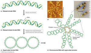
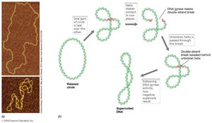
Size, Shape, and Supercoiling: DNA molecules are typically very large and must be compacted to fit within the cell. Supercoiling is a process that helps in this compaction.
Topoisomerases: Enzymes that insert and remove supercoils in DNA, maintaining its structural integrity.
Negative Supercoiling: DNA is twisted in the opposite sense relative to the right-handed double helix, commonly found in most cells.
DNA Gyrase: Introduces negative supercoils into DNA via double-strand breaks.
Positive Supercoiling: Helps prevent DNA melting at high temperatures, important for thermophilic organisms.
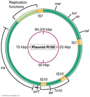
Genetic Elements: Chromosomes and Plasmids
Microbial genomes are composed of chromosomes and plasmids, each with distinct structural and functional properties.
Chromosomes: Main genetic element in cells, usually a single circular DNA molecule in bacteria.
Plasmids: Double-stranded DNA molecules that replicate independently from the chromosome. They are usually circular and often carry genes beneficial to the cell, such as antibiotic resistance.
Viruses: Contain either RNA or DNA genomes, which can be linear or circular, single- or double-stranded.
Extracellular vs. Intracellular: Plasmids are not extracellular, unlike viruses.

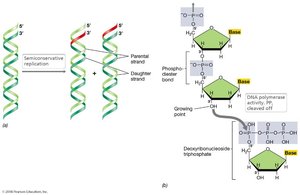
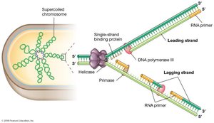
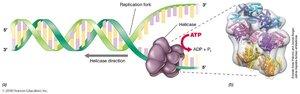
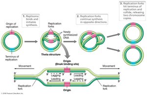
DNA Replication
DNA replication is a highly regulated process that ensures the accurate duplication of genetic material before cell division.
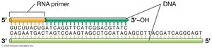
Semiconservative Replication: Each new DNA molecule consists of one parental and one newly synthesized strand.
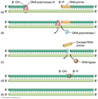
DNA Polymerase: Enzyme responsible for synthesizing new DNA strands by adding nucleotides to the growing chain.
RNA Primers: Short RNA sequences required to initiate DNA synthesis.
Leading and Lagging Strands: DNA is synthesized continuously on the leading strand and discontinuously on the lagging strand (Okazaki fragments).
Molecular Information Flow: Replication, Transcription, Translation
The central dogma of molecular biology describes the flow of genetic information from DNA to RNA to protein. Each step is catalyzed by specific enzymes and involves distinct molecular mechanisms.
Replication: DNA is copied to produce identical genetic material for daughter cells.
Transcription: DNA is used as a template to synthesize RNA.
Translation: mRNA is used as a template to synthesize proteins.
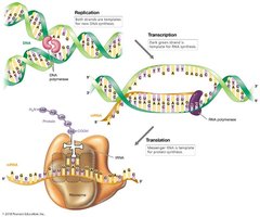
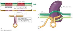
Transcription in Bacteria
Transcription is the process by which RNA is synthesized from a DNA template. In bacteria, this process is regulated by specific DNA sequences and proteins.
Termination of RNA Synthesis: Governed by specific DNA sequences, such as GC-rich regions with inverted repeats.
Rho-dependent Termination: Rho protein recognizes specific DNA sequences, causing RNA polymerase to pause and release the RNA transcript.
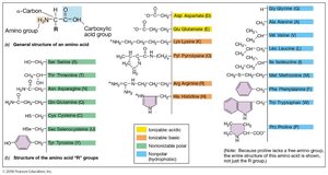
Amino Acids, Polypeptides, and Proteins
Proteins are essential macromolecules in cells, serving catalytic, structural, and regulatory functions. They are polymers of amino acids linked by peptide bonds.
Catalytic Proteins: Enzymes that accelerate biochemical reactions.
Structural Proteins: Components of membranes, cell walls, and ribosomes.
Regulatory Proteins: Control cellular processes and gene expression.
Polypeptides: Chains of amino acids; proteins may consist of one or more polypeptides.
Translation and the Genetic Code
Translation is the process by which proteins are synthesized from mRNA. The genetic code is a set of rules by which nucleotide sequences are translated into amino acid sequences.
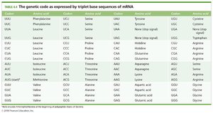
Genetic Code: A triplet of nucleic acid bases (codon) encodes a single amino acid. There are 64 possible codons.
Start and Stop Codons: Specific codons signal the initiation and termination of translation.
Degenerate Code: Multiple codons encode a single amino acid.
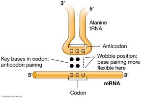
Wobble: Irregular base pairing allowed at the third position of tRNA, increasing flexibility in codon recognition.
Codon | Amino Acid |
|---|---|
UUU, UUC | Phenylalanine |
UUA, UUG, CUU, CUC, CUA, CUG | Leucine |
AUG | Methionine (Start) |
UAA, UAG, UGA | Stop |
... (see full table in image_20) | ... |
Translation
Translation occurs at the ribosome, where mRNA is decoded to synthesize polypeptides. In bacteria and archaea, transcription and translation are often coupled.
Direction of Synthesis: N-terminal to C-terminal.
Polyribosome: Complex of mRNA with several ribosomes, allowing simultaneous translation.
Transfer RNA (tRNA)
tRNA molecules are essential for translation, carrying amino acids to the ribosome and matching them to the appropriate codons in mRNA.
Tertiary Structure: Due to base pairing within the tRNA molecule.
Anticodon: Complementary to the mRNA codon, located on the anticodon arm.
Amino Acid Activation
Amino acids are attached to tRNA molecules by aminoacyl-tRNA synthetases, ensuring the correct pairing of amino acid and tRNA.
Specificity: Each synthetase is specific for a single amino acid and its cognate tRNAs.
The Ribosome
The ribosome is the site of protein synthesis, composed of two subunits in bacteria: 30S and 50S, forming the 70S ribosome.
Translational Domain: Responsible for translation on both subunits.
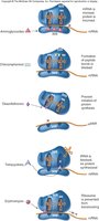
Elongation of the Polypeptide Chain
Elongation involves the sequential addition of amino acids to the growing polypeptide chain, facilitated by elongation factors.
Aminoacyl-tRNA Binding
Transpeptidation Reaction
Translocation
tRNA Binding Sites of Ribosome
The ribosome contains three binding sites for tRNA:
Peptidyl (P) Site: Binds initiator tRNA or tRNA attached to growing polypeptide.
Aminoacyl (A) Site: Binds incoming aminoacyl-tRNA.
Exit (E) Site: Briefly binds empty tRNA before it leaves the ribosome.
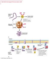
Assisted Protein Folding and Chaperones
Chaperones are proteins that assist in the proper folding of other proteins, preventing misfolding and aggregation.
DnaK, DnaJ, GroEL, GroES: Key chaperones in Escherichia coli.
ATP-dependent: DnaK and DnaJ slow polypeptide folding; GroEL and GroES fold partially folded proteins.
Refolding: Chaperones can refold partially denatured proteins.
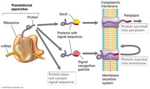
Protein Secretion: The Sec and Tat Systems
Protein secretion systems transport proteins across bacterial and archaeal membranes. The Sec system exports unfolded proteins, while the Tat system transports folded proteins.
Signal Sequence: 15–20 residues at the N-terminus signal the secretory system and prevent complete folding.
Antibiotics that Inhibit Bacterial Translation
Several antibiotics target bacterial translation, interfering with protein synthesis and thus inhibiting bacterial growth.
Aminoglycosides (Gentamicin, Streptomycin): Distort ribosome shape.
Chloramphenicol: Prevents peptide bond formation.
Tetracycline: Blocks tRNA attachment to mRNA.
Erythromycin: Prevents the shift on the ribosome.

Additional info: Academic context and explanations have been expanded for clarity and completeness. Table 4.4 is partially recreated; see image_20 for full codon-to-amino acid mapping.
