 Back
BackMicrobial Growth and Cell Division: Mechanisms and Regulation
Study Guide - Smart Notes
 Tailored notes based on your materials, expanded with key definitions, examples, and context.
Microbial Growth and Cell Division
Introduction to Microbial Growth
Microbial growth refers to the increase in the number of cells in a population, primarily through cell division. This process is fundamental to the propagation of bacteria, archaea, and microbial eukaryotes. Understanding the mechanisms of cell division and the regulation of the cell cycle is essential for microbiology students, as these processes underpin microbial physiology, genetics, and ecology.
Eukaryotic Cell Cycle
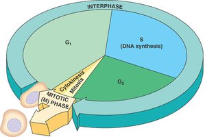
Phases of the Eukaryotic Cell Cycle
The eukaryotic cell cycle is divided into interphase (G1, S, G2) and the mitotic (M) phase, which includes mitosis and cytokinesis. Interphase is the period of cell growth and DNA replication, while the M phase is where cell division occurs.
G1 phase: Cell growth and preparation for DNA synthesis.
S phase: DNA synthesis (replication) occurs.
G2 phase: Further growth and preparation for mitosis.
M phase: Mitosis (nuclear division) and cytokinesis (cytoplasmic division).
Bacterial Cell Cycle and Binary Fission
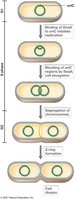
Overview of the Bacterial Cell Cycle
Bacteria typically reproduce by binary fission, a process that involves the replication of the chromosome, segregation of the genetic material, and division of the cell into two genetically identical daughter cells.
DNA replication: Begins at the origin of replication and proceeds bidirectionally.
Chromosome segregation: Each daughter cell receives one copy of the chromosome.
Divisome formation: Protein complex assembles at the future site of division.
Cell elongation and septum formation: The cell elongates and a septum forms, dividing the cell.
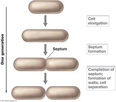
Binary Fission Process
Binary fission is the most common mode of bacterial reproduction. It involves cell elongation, septum formation, and separation into two cells.
Cell elongation: The cell grows in size.
Septum formation: A partition forms between the two future daughter cells.
Cell separation: The septum is completed, and the cells separate.
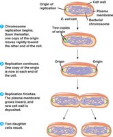
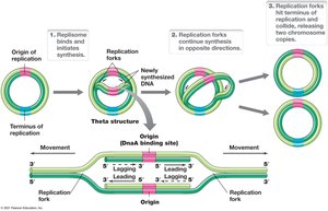
Chromosome Replication and Partitioning in Bacteria
During binary fission, the bacterial chromosome is replicated and partitioned so that each daughter cell receives a complete copy. The process is tightly regulated to ensure genetic fidelity.
Origin of replication (oriC): Site where DNA replication begins.
Bidirectional replication: Two replication forks move in opposite directions around the circular chromosome.
Partitioning: The origins move to opposite ends of the cell, and the cell divides.
Bacterial Chromosomes and Plasmids
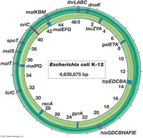
Structure of the Bacterial Chromosome
Bacterial chromosomes are typically circular DNA molecules containing essential genes for survival. The Escherichia coli K-12 chromosome is a well-studied example, with a size of approximately 4.6 million base pairs.
Genetic linkage map: Shows the relative positions of genes on the chromosome.
oriC: The origin of replication is a key feature.
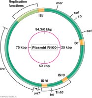
Bacterial Plasmids
Plasmids are extrachromosomal DNA elements that often carry genes for antibiotic resistance, virulence, or metabolic functions. They replicate independently of the chromosome.
Resistance plasmids (R plasmids): Carry genes that confer resistance to antibiotics.
Replication functions: Plasmids have their own origins of replication and regulatory elements.
DNA Structure and Packaging
DNA Structure
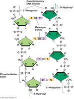
DNA is a double-stranded helix composed of nucleotides. Each nucleotide consists of a deoxyribose sugar, a phosphate group, and a nitrogenous base (adenine, thymine, cytosine, or guanine). The strands are held together by hydrogen bonds between complementary bases.
Phosphodiester bonds: Link the sugar-phosphate backbone.
Base pairing: A pairs with T, and G pairs with C.
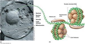
DNA Packaging in Microorganisms
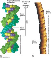
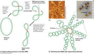
DNA must be compacted to fit within the cell. Bacteria and archaea use supercoiling, while eukaryotes use histones to form nucleosomes.
Supercoiling: Introduced by DNA gyrase in bacteria, compacts the DNA.
Nucleoid: The region in bacteria where the chromosome is located.
Nucleosomes: In eukaryotes, DNA wraps around histone proteins.
DNA Replication in Bacteria
Initiation of Replication
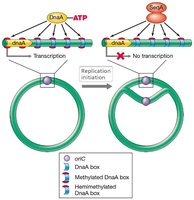
DNA replication begins at the origin of replication (oriC) and is regulated by initiator proteins such as DnaA. The process is tightly controlled to ensure that replication occurs only once per cell cycle.
DnaA: Binds to oriC and initiates unwinding of the DNA.
Regulation: Methylation and other factors control the timing of initiation.
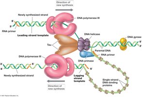
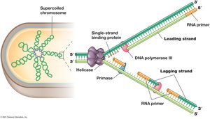
Replication Fork and the Replisome
The replisome is a complex of enzymes that carries out DNA replication at the replication fork. Key enzymes include DNA polymerase III, helicase, primase, and DNA gyrase.
Leading strand: Synthesized continuously in the 5' to 3' direction.
Lagging strand: Synthesized discontinuously as Okazaki fragments.
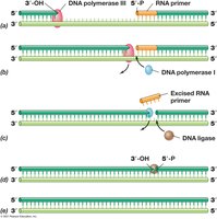
Events at the Replication Fork
At the replication fork, the DNA is unwound, and new strands are synthesized. RNA primers are required to initiate synthesis, and DNA ligase seals nicks in the lagging strand.
Primase: Synthesizes short RNA primers.
DNA polymerase III: Extends the primers with DNA.
DNA polymerase I: Removes RNA primers and fills in with DNA.
DNA ligase: Seals the nicks between Okazaki fragments.
Termination of Replication
Replication of circular chromosomes ends at specific termination sites (Ter), where proteins such as Tus block the replication fork. In eukaryotes, telomerase completes the ends of linear chromosomes.
Theta Structure in Circular DNA Replication
During replication of circular DNA, the intermediate structure resembles the Greek letter theta (θ), reflecting the bidirectional movement of replication forks.
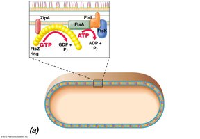
Bacterial Cell Division Machinery
Divisome and Fts Proteins
The divisome is a protein complex that orchestrates bacterial cell division. Key proteins include FtsZ, which forms a ring at the future division site, and FtsA, which anchors FtsZ to the membrane. Min proteins ensure the divisome forms at the cell center.
FtsZ: Tubulin-like protein that forms the Z ring.
FtsA: Connects FtsZ to the membrane.
Min proteins: Prevent division at the cell poles.
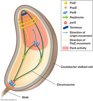
Chromosome Partitioning in Caulobacter
Some bacteria, such as Caulobacter, have specialized mechanisms for chromosome partitioning involving proteins like ParA and PopZ, ensuring accurate segregation during cell division.
Peptidoglycan Synthesis and Cell Wall Expansion
Structure of Peptidoglycan
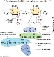
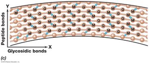
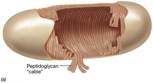
Peptidoglycan is a polymer consisting of sugars (N-acetylglucosamine and N-acetylmuramic acid) and peptides. It provides structural integrity to the bacterial cell wall.
Glycan tetrapeptide: The repeating unit of peptidoglycan.
Crosslinks: Peptide bonds connect glycan chains, forming a mesh-like structure.
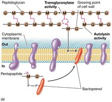
Peptidoglycan Synthesis
Peptidoglycan synthesis involves the formation of precursors, their transport across the membrane by bactoprenol, and their insertion into the existing cell wall by transglycosylases and transpeptidases (e.g., FtsI).
Autolysins: Create openings in the cell wall for new material.
Bactoprenol: Lipid carrier that transports precursors.
Transpeptidation: Formation of peptide crosslinks.

Patterns of Microbial Growth
Growth Phases in Batch Culture
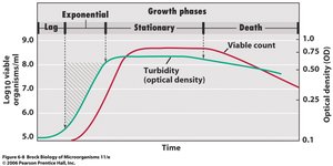
Microbial populations in batch culture exhibit distinct growth phases: lag, exponential (log), stationary, and death. These phases reflect changes in nutrient availability and waste accumulation.
Lag phase: Adaptation and enzyme synthesis.
Exponential phase: Rapid, constant cell division.
Stationary phase: Growth rate slows as nutrients deplete.
Death phase: Cells die due to harsh conditions.
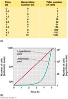
Mathematics of Exponential Growth
Exponential growth can be described mathematically, allowing prediction of cell numbers over time. The generation time (g) is the time required for the population to double.
Key equations:
N: Final number of cells
N0: Initial number of cells
n: Number of generations
g: Generation time
t: Time of exponential growth
Batch vs. Continuous Culture
Batch cultures are closed systems with fixed nutrients, while continuous cultures (chemostats) maintain cells in exponential growth by continuously adding fresh medium and removing waste.
Batch culture: Used for routine laboratory studies.
Chemostat: Allows precise control of growth rate and cell density.
Summary Table: Key Proteins in Bacterial Cell Division
Protein | Function |
|---|---|
FtsZ | Forms the Z ring at the division site |
FtsA | Anchors FtsZ to the membrane |
Min proteins | Ensure division occurs at midcell |
FtsI | Transpeptidase for peptidoglycan synthesis |
MreB | Determines cell shape by directing cell wall synthesis |
Additional info: This guide integrates foundational concepts from microbial cell structure, function, and growth, as well as the molecular mechanisms of DNA replication and cell division, as outlined in standard microbiology curricula.
