 Back
BackMicrobial Growth and Cell Division: Structure, Function, and Regulation
Study Guide - Smart Notes
 Tailored notes based on your materials, expanded with key definitions, examples, and context.
Microbial Growth and Cell Division
Overview of Microbial Growth
Microbial growth refers to the increase in the number of cells in a population, rather than an increase in cell size. Most bacteria and archaea reproduce by binary fission, while eukaryotic microorganisms may reproduce by mitosis, budding, or other mechanisms. Understanding the cell cycle and division processes is essential for studying microbial physiology and genetics.
Eukaryotic Cell Cycle
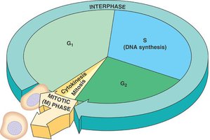
Phases of the Eukaryotic Cell Cycle
The eukaryotic cell cycle is divided into interphase (G1, S, G2) and the mitotic (M) phase. Interphase is the period of cell growth and DNA replication, while the M phase includes mitosis and cytokinesis.
G1 phase: Cell growth and preparation for DNA synthesis.
S phase: DNA synthesis (replication) occurs.
G2 phase: Further growth and preparation for mitosis.
M phase: Mitosis (nuclear division) and cytokinesis (cytoplasmic division).
Bacterial Cell Cycle and Division
Overview of the Bacterial Cell Cycle
Bacteria typically reproduce by binary fission, a process involving cell growth, DNA replication, chromosome segregation, and division. The cell cycle is tightly regulated to ensure accurate genetic inheritance.
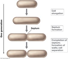
Binary Fission
Binary fission is the most common form of cell division in prokaryotes. It involves elongation of the cell, replication of the chromosome, formation of a septum, and separation into two daughter cells.
Cell elongation: The cell increases in size as it prepares to divide.
Septum formation: A partition (septum) forms, dividing the cell into two compartments.
Cell separation: The septum is completed, and the two daughter cells separate.
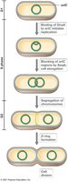
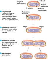
Chromosome Replication and Partitioning
During binary fission, the bacterial chromosome is replicated, and each copy is segregated to opposite ends of the cell. The cell then divides, ensuring each daughter cell receives a complete genome.
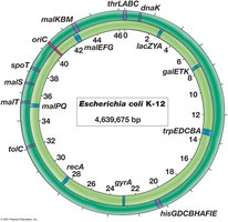
Origin of replication (oriC): The site where DNA replication begins.
Replication forks: Structures that move bidirectionally from the origin, synthesizing new DNA.
Partitioning: The two chromosomes are actively separated before cell division.
Bacterial Chromosomes and Plasmids
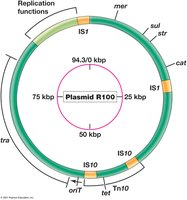
Bacterial genomes are typically composed of a single, circular chromosome. Many bacteria also contain plasmids—small, circular DNA molecules that replicate independently and often carry genes for antibiotic resistance or other functions.
Chromosome: Contains essential genes for cell function and replication.
Plasmid: May carry accessory genes, such as those conferring resistance to antibiotics.
DNA Structure and Packaging
Structure of DNA
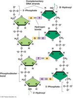
DNA is a double-stranded helix composed of nucleotides. Each nucleotide consists of a deoxyribose sugar, a phosphate group, and a nitrogenous base (adenine, thymine, cytosine, or guanine). The two strands are held together by hydrogen bonds between complementary bases.
Phosphodiester bonds: Link the sugar-phosphate backbone of each strand.
Base pairing: Adenine pairs with thymine (A=T), and cytosine pairs with guanine (C≡G).
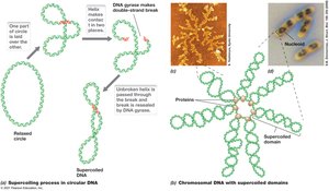
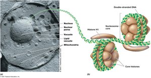
DNA Supercoiling and Packaging
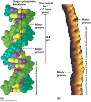
To fit within the small volume of a cell, DNA is highly compacted. In bacteria, DNA is supercoiled by enzymes such as DNA gyrase. In eukaryotes, DNA is wrapped around histone proteins to form nucleosomes.
Supercoiling: Introduces twists into the DNA molecule, reducing its effective length.
DNA gyrase: An enzyme that introduces negative supercoils into DNA.
Nucleoid: The region in a prokaryotic cell where the chromosome is located.
Nucleosome: The basic unit of DNA packaging in eukaryotes.
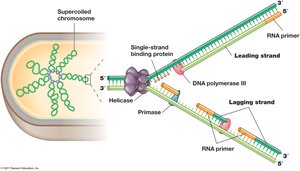
DNA Replication in Bacteria
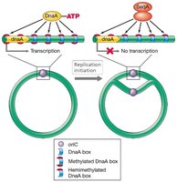
Initiation of Replication
DNA replication begins at the origin of replication (oriC) and is regulated by initiator proteins such as DnaA. The process is tightly controlled to ensure replication occurs only once per cell cycle.
DnaA: Binds to the origin and facilitates unwinding of DNA.
Regulation: Methylation and other factors control the timing of initiation.
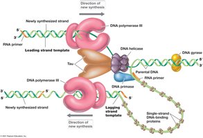
Replication Fork and the Replisome
The replication fork is the site where DNA is unwound and new strands are synthesized. The replisome is a complex of enzymes responsible for DNA replication, including DNA polymerase, helicase, primase, and others.
Leading strand: Synthesized continuously in the 5' to 3' direction.
Lagging strand: Synthesized discontinuously as Okazaki fragments.
DNA ligase: Seals nicks between Okazaki fragments.
Termination of Replication
In bacteria with circular chromosomes, replication terminates at specific Ter sites, where Tus proteins block the replication fork. Eukaryotes use telomerase to complete the ends of linear chromosomes.
Ter sites: DNA sequences that signal the end of replication.
Tus protein: Binds Ter sites and halts the replication machinery.
Telomerase: Enzyme that extends telomeres in eukaryotic chromosomes.
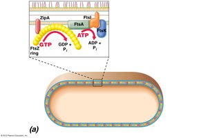
Cell Division Machinery in Bacteria
The Divisome and Fts Proteins
The divisome is a protein complex that mediates bacterial cell division. Key proteins include FtsZ, which forms a ring at the future site of division, and FtsA, which anchors FtsZ to the membrane. Min proteins ensure the divisome forms at the cell center.
FtsZ: Tubulin-like protein that assembles into a ring at the division site.
FtsA: Connects FtsZ to the cytoplasmic membrane.
Min proteins: Prevent formation of the divisome at cell poles.
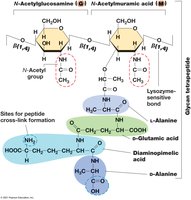
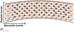
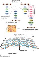
Cell Wall Synthesis and Morphology
During division, new peptidoglycan is inserted into the cell wall. MreB proteins help determine cell shape by directing cell wall synthesis, while FtsI (a penicillin-binding protein) catalyzes transpeptidation, forming peptide cross-links in peptidoglycan.
Autolysins: Enzymes that create openings in the cell wall for new material insertion.
Bactoprenol: Lipid carrier that transports peptidoglycan precursors across the membrane.
Transglycosylases: Form glycosidic bonds between sugars in peptidoglycan.
Transpeptidases: Form peptide cross-links between peptidoglycan chains.
Patterns of Microbial Growth
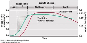
Growth Phases in Batch Culture
When microorganisms are inoculated into a fresh medium, they exhibit a characteristic growth curve with four phases: lag, exponential (log), stationary, and death.
Lag phase: Cells adapt to new conditions; little or no cell division.
Exponential phase: Cells divide at a constant, maximum rate.
Stationary phase: Growth rate slows as nutrients are depleted and waste accumulates.
Death phase: Cells die due to unfavorable conditions.
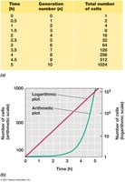
Mathematics of Exponential Growth
Microbial populations grow exponentially during the log phase. The number of cells at any time can be calculated using the following equations:
Exponential growth equation:
Generation time (g):
Where: N = final cell number, N0 = initial cell number, n = number of generations, t = time of exponential growth.
Batch vs. Continuous Culture
Microbial cultures can be grown in batch or continuous systems. Batch cultures are closed systems with a fixed volume, while continuous cultures (e.g., chemostats) maintain cells in exponential growth by continuously adding fresh medium and removing waste.
Batch culture: Used for routine laboratory studies; exhibits all growth phases.
Chemostat: Allows precise control of growth rate and cell density.
Summary Table: Key Proteins and Structures in Bacterial Cell Division
Protein/Structure | Function |
|---|---|
FtsZ | Forms ring at division site; initiates septum formation |
FtsA | Anchors FtsZ to membrane |
Min proteins | Ensure division occurs at cell center |
MreB | Determines cell shape; directs cell wall synthesis |
FtsI | Transpeptidase; catalyzes cross-linking in peptidoglycan |
Bactoprenol | Transports peptidoglycan precursors across membrane |
Autolysins | Create openings in cell wall for new material insertion |
Conclusion
Understanding microbial growth and cell division is fundamental to microbiology. The processes of DNA replication, chromosome segregation, and cell wall synthesis are tightly coordinated to ensure the survival and propagation of microbial populations. These mechanisms are targets for antibiotics and are central to microbial physiology, genetics, and biotechnology.
