 Back
BackMicrobial Growth: Environmental and Nutritional Factors, Cultivation, and Measurement
Study Guide - Smart Notes
 Tailored notes based on your materials, expanded with key definitions, examples, and context.
Microbial Growth
Biofilms
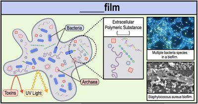
Biofilms are structured communities of microorganisms encased in a self-produced matrix of extracellular polymeric substances (EPS) and attached to a surface. These communities can include bacteria and archaea and are commonly found in natural, industrial, and clinical settings.
Definition: Biofilms are organized assemblies of microbial cells surrounded by a sticky, slime-like EPS layer anchored to a surface.
EPS (Extracellular Polymeric Substance): The EPS matrix is secreted by cells and provides structural support, protection from harmful conditions (e.g., UV light, toxins, antibiotics), and facilitates the exchange of genetic material.
Genetic Exchange: Resistance genes can be transferred between organisms via DNA within the EPS, enhancing survival under stress.
Quorum Sensing: Cells within a biofilm communicate chemically to coordinate behavior based on population density.
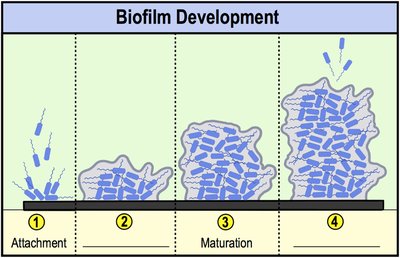
Biofilm Development
Biofilm formation occurs in four main stages:
Attachment: Cells adhere to a surface, often using fimbriae.
Colonization: Cells multiply and produce EPS.
Maturation: Additional cells attach and multiply as the EPS matrix expands.
Dispersal: Cells detach to colonize new environments.
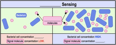
Quorum Sensing
Quorum sensing is a process by which bacteria detect their population density through the accumulation of signaling molecules in the EPS. When the concentration of these molecules reaches a threshold, it triggers coordinated gene expression across the community.
Low cell density = low signal molecule concentration.
High cell density = high signal molecule concentration, activating group behaviors.
Growing a Pure Culture
Pure Culture and Colony Formation
In laboratory settings, microbiologists isolate and grow microorganisms in pure cultures to study their properties.
Pure Culture: A population of cells derived from a single parent cell, containing only one species.
Colony: A visible mass of cells (about 1 million) originating from a single cell.
Inoculum: The sample of cells introduced into a culture medium to initiate growth.

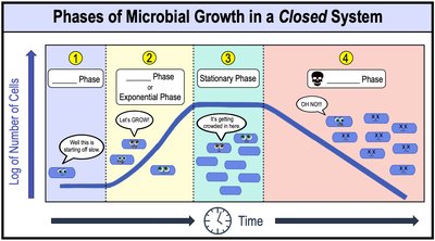
Microbial Growth Curves in a Closed System
Phases of Growth
When microbes are grown in a closed system (batch culture), their population follows a characteristic growth curve with four phases:
Lag Phase: Cells adapt to the environment and synthesize necessary enzymes; little to no cell division occurs.
Log (Exponential) Phase: Cells divide at a constant, rapid rate; population increases logarithmically.
Stationary Phase: Nutrient depletion and waste accumulation halt growth; cell division rate equals death rate.
Death Phase: Cells die at an exponential rate due to lack of nutrients and toxic conditions.
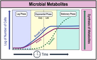
Microbial Metabolites
Microbes produce different metabolites during various growth phases:
Primary Metabolites: Produced during early log phase; essential for growth (e.g., amino acids, nucleotides).
Secondary Metabolites: Produced during late log and stationary phases; not required for growth but may provide survival advantages (e.g., antibiotics).
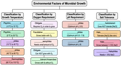
Environmental Factors Affecting Microbial Growth
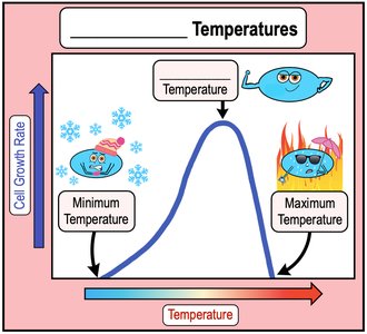
Temperature Requirements
Microbial species have specific temperature ranges for growth, defined by minimum, optimum, and maximum cardinal temperatures.
Minimum Temperature: Lowest temperature for growth; below this, growth ceases.
Optimum Temperature: Temperature at which growth rate is highest.
Maximum Temperature: Highest temperature for growth; above this, proteins denature and cells die.
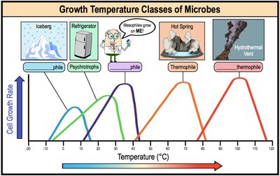
Classification by Growth Temperature
Psychrophiles: -5°C to 20°C (cold-loving)
Psychrotrophs: 0°C to 35°C (can grow at refrigeration temperatures)
Mesophiles: 10°C to 45°C (moderate temperatures; includes most human pathogens)
Thermophiles: 40°C to 80°C (heat-loving)
Hyperthermophiles: 65°C to 115°C (extreme heat-loving)
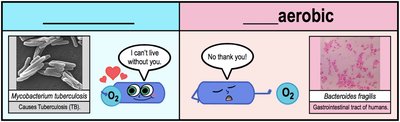
Oxygen Requirements
Microbes are classified based on their oxygen requirements for growth:
Obligate Aerobes: Require O2 for growth.
Obligate Anaerobes: Cannot grow in the presence of O2.
Facultative Anaerobes: Grow better with O2 but can grow without it.
Microaerophiles: Require low levels of O2.
Aerotolerant Anaerobes: Indifferent to O2; do not use it but can tolerate its presence.
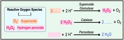
Reactive Oxygen Species (ROS) and Enzymes
Aerobic metabolism produces toxic ROS, which are neutralized by enzymes:
Superoxide Dismutase (SOD): Converts superoxide radicals to hydrogen peroxide and oxygen.
Catalase: Converts hydrogen peroxide to water and oxygen.
Peroxidase: Converts hydrogen peroxide to water.
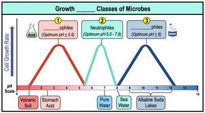
pH Requirements
Microbes grow within specific pH ranges and are classified by their optimal pH:
Acidophiles: Optimum pH < 5.5 (e.g., microbes in volcanic soil, stomach acid)
Neutrophiles: Optimum pH 5.5–7.9 (e.g., most human pathogens)
Alkaliphiles: Optimum pH > 8 (e.g., microbes in alkaline lakes)
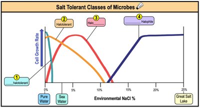
Osmolarity and Salt Tolerance
Microbes are also classified by their tolerance to salt concentrations:
Non-halotolerant: Cannot tolerate moderate salt concentrations.
Halotolerant: Can tolerate moderate salt concentrations (e.g., skin bacteria).
Halophiles: Require 1–14% NaCl (e.g., marine bacteria).
Extreme Halophiles: Require >15% NaCl (e.g., Great Salt Lake bacteria).
Summary Table: Environmental Factors of Microbial Growth
Nutritional Factors of Microbial Growth
Energy, Electron, and Carbon Sources
Microbes are classified by their sources of energy, electrons, and carbon:
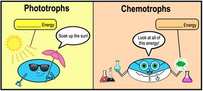
Energy Source:
Phototrophs: Obtain energy from sunlight.
Chemotrophs: Obtain energy from chemical compounds.
Electron Source:
Lithotrophs: Use reduced inorganic molecules (e.g., H2O, Fe2+).
Organotrophs: Use organic molecules (e.g., glucose).
Carbon Source:
Autotrophs: Fix inorganic CO2 to synthesize organic molecules.
Heterotrophs: Use preformed organic molecules as a carbon source.
Summary Table: Nutritional Classifications
Energy Source | Electron Source | Carbon Source |
|---|---|---|
Phototroph: Sunlight | Lithotroph: Inorganic molecules | Autotroph: CO2 |
Chemotroph: Chemical compounds | Organotroph: Organic molecules | Heterotroph: Organic molecules |

Cultivating Microbial Growth
Culture Media
Microbes are grown in the laboratory using culture media, which can be solid (agar) or liquid (broth). Aseptic technique is essential to prevent contamination.
Agar: A polysaccharide from marine algae used to solidify media.
Types of Solid Media: Slants (agar in tubes at an angle), deeps (agar solidified upright), and Petri dishes (plates).
Types of Culture Media
Chemically Defined Media: Exact chemical composition is known; used for precise experiments.
Complex Media: Contains extracts from yeast, meat, or plants; composition varies.
Selective Media: Promotes growth of specific microbes while inhibiting others (e.g., MacConkey agar).
Differential Media: Contains indicators to distinguish between species based on biochemical reactions (e.g., blood agar for hemolysis).
Reducing Media: Contains agents to remove oxygen for growing anaerobes.
Enrichment Media: Favors growth of a particular microbe present in low numbers without inhibitors.
Measuring Microbial Growth
Direct Cell Counts
Direct Microscopic Count: Cells are counted under a microscope using a counting chamber; does not distinguish live from dead cells.
Flow Cytometry: Cells pass through a laser beam and are counted electronically.
Coulter Counter: Electronically counts cells as they pass through a channel.
Viable Plate Counts
Plate Count: Only viable cells capable of forming colonies are counted; requires serial dilution for accuracy.
Colony Forming Unit (CFU): Each colony represents a viable cell from the original sample.
Membrane Filtration
Used for samples with low cell numbers; cells are trapped on a membrane filter, then transferred to agar for colony counting.
Measuring Biomass
Spectrophotometry: Measures turbidity (cloudiness) of a culture; higher turbidity indicates higher biomass.
Total Weight: Cells are centrifuged, and the pellet is weighed.
Summary Table: Types of Culture Media
Type | Description | Example |
|---|---|---|
Chemically Defined | Exact composition known | Minimal media |
Complex | Contains extracts, composition varies | Nutrient broth |
Selective | Inhibits unwanted microbes | MacConkey agar |
Differential | Distinguishes species visually | Blood agar |
Reducing | Removes oxygen | Thioglycolate broth |
Enrichment | Favors specific microbe | Selenite broth |
