Back
BackMicrobial Mechanisms of Pathogenicity and Disease Processes
Study Guide - Smart Notes
 Tailored notes based on your materials, expanded with key definitions, examples, and context.
Disease Processes: Microbial Mechanisms of Pathogenicity
Overview of Pathogenic Mechanisms
Pathogenic microorganisms cause disease through a series of well-defined steps, including entry into the host, adherence to host tissues, evasion of host defenses, and damage to host cells. Understanding these mechanisms is crucial for the study of infectious diseases in microbiology.
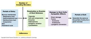
Portals of Entry: Microbes enter the host through specific routes such as mucous membranes, skin, and the parenteral route (direct deposition beneath the skin).
Adherence: Pathogens attach to host cells using adhesion factors, which are often proteins or glycoproteins on the microbial surface.
Penetration/Evasion of Host Defenses: Microbes use capsules, cell wall components, and enzymes to evade or overcome host immune responses.
Damage to Host Cells: Pathogens may cause direct damage, produce toxins (exotoxins and endotoxins), or induce cytopathic effects.
Portals of Exit: Microbes leave the host via routes similar to their entry, facilitating transmission to new hosts.
Portals of Entry and Exit
Major Routes of Entry and Exit
Microorganisms gain access to the body through several portals, which are also commonly used as exit routes to spread infection.
Mucous membranes: Respiratory, gastrointestinal, and genitourinary tracts are common entry points.
Skin: Although an effective barrier, some pathogens can enter through cuts or abrasions.
Parenteral route: Direct introduction into tissues, such as via punctures, bites, or injections.
Portals of exit are generally the same as the portals of entry, allowing pathogens to spread to new hosts.
Adhesion Factors
Mechanisms of Microbial Attachment
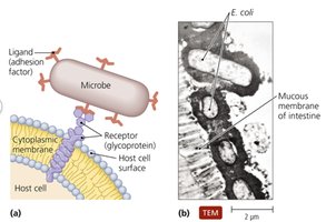
Adhesion is a critical step in the establishment of infection. Microbes use specialized structures or molecules called adhesins to bind to specific receptors on host cells.
Ligands (adhesion factors): Surface molecules on pathogens that bind to complementary receptors on host cells.
Host cell receptors: Usually glycoproteins or glycolipids on the host cell membrane.
Specificity: The interaction between adhesins and receptors determines host and tissue specificity.
Virulence Factors: Extracellular Enzymes
Role of Enzymes in Pathogenicity
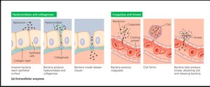
Many pathogens secrete enzymes that facilitate invasion and damage to host tissues. These enzymes can degrade host barriers or interfere with immune responses.
Hyaluronidase and Collagenase: Break down connective tissue components, allowing deeper penetration of bacteria.
Coagulase and Kinase: Coagulase promotes clotting to protect bacteria, while kinase dissolves clots to release bacteria.
Bacterial Toxins: Exotoxins and Endotoxins
Types and Actions of Bacterial Toxins
Bacterial toxins are major contributors to disease symptoms. They are classified as exotoxins or endotoxins based on their origin and properties.
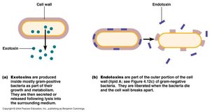
Exotoxins: Proteins secreted by bacteria (mainly Gram-positive) that cause specific effects in the host.
Endotoxins: Lipopolysaccharide (LPS) components of the outer membrane of Gram-negative bacteria, released upon cell death.
Comparison of Exotoxins and Endotoxins
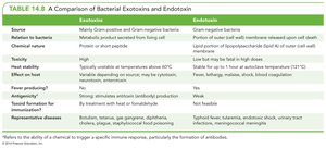
The following table summarizes the key differences between exotoxins and endotoxins:
Feature | Exotoxins | Endotoxins |
|---|---|---|
Source | Mainly Gram-positive and some Gram-negative bacteria | Gram-negative bacteria |
Chemical Nature | Protein, usually with enzymatic activity | Lipid A portion of lipopolysaccharide (LPS) |
Toxicity | High | Low (but can be fatal in high doses) |
Heat Stability | Unstable, inactivated at 60°C | Stable at 121°C |
Fever Producing | No | Yes |
Antigenicity | Strong, stimulates antibody production | Weak |
Representative Diseases | Botulism, tetanus, diphtheria | Typhoid fever, meningococcemia |
Action of Gram-Negative Endotoxin (LPS)
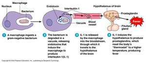
Endotoxins, specifically the lipid A component of LPS, trigger strong immune responses, including fever and inflammation. The process involves macrophage activation and release of cytokines such as interleukin-1 (IL-1), which acts on the hypothalamus to induce fever.
Step 1: Macrophage ingests Gram-negative bacterium.
Step 2: Bacterium is degraded, releasing endotoxin.
Step 3: Endotoxin stimulates macrophage to release IL-1.
Step 4: IL-1 travels to the hypothalamus, inducing prostaglandin production and fever.
Types of Exotoxins
Classification and Examples
Exotoxins are classified based on their target and mechanism of action:
Neurotoxins: Affect nerve cells (e.g., Clostridium botulinum and C. tetani neurotoxins).
Enterotoxins: Affect the gastrointestinal tract (e.g., Staphylococcus aureus and Vibrio cholerae enterotoxins).
Cytotoxins: Kill or damage host cells (e.g., Corynebacterium diphtheriae and Bacillus anthracis cytotoxins).
Exotoxin genes may be located on the bacterial chromosome, plasmids, or lysogenic bacteriophages.
Infection versus Intoxication
Key Differences
It is important to distinguish between infection and intoxication in microbial diseases:
Infection: Involves the multiplication of the microorganism in the host, with an incubation period of at least 12 hours. The immune response is often observed, and toxins may or may not be involved.
Intoxication: Results from ingestion of pre-formed exotoxins. The incubation period is shorter (up to 5-6 hours), the microorganism may not be present in the host, and there is no immune response. Exotoxins are always involved.
Examples of Diseases Caused by Neurotoxins
Tetanus – Infection
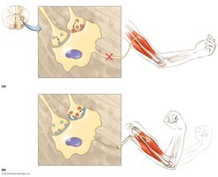
Clostridium tetani produces tetanospasmin, a neurotoxin that blocks the release of inhibitory neurotransmitters, resulting in spastic paralysis. The infection is typically acquired through deep puncture wounds, where anaerobic conditions allow bacterial growth and toxin production.
Mechanism: Tetanospasmin prevents muscle relaxation by inhibiting inhibitory neurotransmitter release in the spinal cord.
Botulism – Intoxication
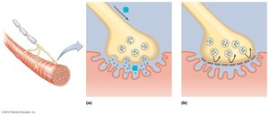
Clostridium botulinum produces botulinum toxin in anaerobic environments, such as improperly canned foods. Ingestion of the toxin leads to flaccid paralysis by blocking acetylcholine release at neuromuscular junctions.
Mechanism: Botulinum toxin prevents muscle contraction by inhibiting neurotransmitter release.
Examples of Diseases Caused by Enterotoxins
Staphylococcal Foodborne Illness – Intoxication
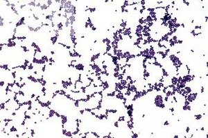
Food contaminated with Staphylococcus aureus can lead to rapid onset of vomiting and gastrointestinal distress due to pre-formed enterotoxins. Temperature abuse of food allows bacterial multiplication and toxin production.
Mechanism: Ingestion of enterotoxin stimulates the GI tract, causing vomiting.
Cholera – Infection
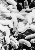
Vibrio cholerae produces cholera toxin in the intestine after ingestion of contaminated water. The toxin causes massive electrolyte and water loss from intestinal cells, resulting in severe diarrhea.
Mechanism: Cholera toxin activates adenylate cyclase, increasing cAMP and causing secretion of Cl–, Na+, and water into the intestinal lumen.
Cytopathic Effect (CPE) of Viruses
Structural Changes in Host Cells
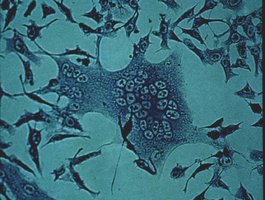
Viruses can induce visible changes in host cells, known as cytopathic effects (CPE), which are useful for diagnosis. These include cell rounding, lysis, syncytia formation (fusion of cells), and inclusion bodies (sites of viral assembly).
Syncytia formation: Fusion of neighboring cells, as seen in measles virus infection.
Inclusion bodies: Aggregates of viral particles or altered host cell components, such as Negri bodies in rabies.
Cell lysis: Loss of cells due to viral replication and destruction.
Effects of Viral Infections Not Seen Under a Light Microscope
Interferon Production and Antigenic Changes
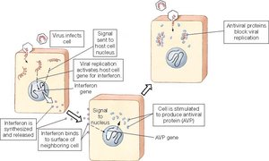
Some effects of viral infection are not visible microscopically but are important for host defense and immune recognition.
Interferon production: Infected cells produce interferons, proteins that inhibit viral replication in neighboring cells.
Antigenic changes: Viral proteins are presented on the surface of infected cells via MHC-I, marking them for immune recognition.
Host-Pathogen Co-Evolution and Disease Severity
Adaptation and Virulence
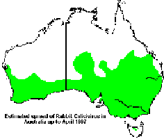
Viruses and their natural hosts often co-evolve, resulting in less severe disease in the natural host. Introduction of a virus into a new host species can lead to high mortality, as seen with the myxoma virus in European rabbits in Australia.
Example: Myxoma virus causes mild disease in South American rabbits (natural host) but severe outbreaks in European rabbits (introduced host).