 Back
BackMicrobial Mechanisms of Pathogenicity and Innate Immunity
Study Guide - Smart Notes
 Tailored notes based on your materials, expanded with key definitions, examples, and context.
Microbial Mechanisms of Pathogenicity
Pathogenicity, Virulence, and Portals of Entry
Pathogenicity refers to the ability of a microorganism to cause disease, while virulence describes the degree of pathogenicity. Microorganisms enter the host through specific portals of entry, which are critical for establishing infection. The main portals include mucous membranes (respiratory, gastrointestinal, genitourinary tracts), skin, and parenteral routes (such as wounds).
Pathogenicity: Ability to cause disease.
Virulence: Degree of pathogenicity.
Portals of Entry: Mucous membranes, skin, parenteral route.
Preferred Portal: Most pathogens have a preferred portal of entry for effective infection.
Portal of Entry | Pathogen | Disease |
|---|---|---|
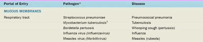
Respiratory tract | Streptococcus pneumoniae | Pneumococcal pneumonia |
Respiratory tract | Mycobacterium tuberculosis | Tuberculosis |
Respiratory tract | Bordetella pertussis | Whooping cough (pertussis) |
Respiratory tract | Influenza virus (Influenzavirus) | Influenza |
Respiratory tract | Measles virus (Morbillivirus) | Measles (rubeola) |
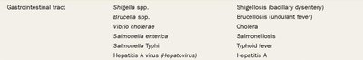
Gastrointestinal tract | Shigella spp. | Shigellosis (bacillary dysentery) |
Gastrointestinal tract | Brucella spp. | Brucellosis (undulant fever) |
Gastrointestinal tract | Vibrio cholerae | Cholera |
Gastrointestinal tract | Salmonella enterica | Salmonellosis |
Gastrointestinal tract | Salmonella Typhi | Typhoid fever |
Gastrointestinal tract | Hepatitis A virus (Hepatovirus) | Hepatitis A |
Genitourinary tract | Neisseria gonorrhoeae | Gonorrhea |
Genitourinary tract | Treponema pallidum | Syphilis |
Genitourinary tract | Chlamydia trachomatis | Nongonococcal urethritis |
Genitourinary tract | Human herpesvirus-2 | Herpes virus infections |
Genitourinary tract | Human immunodeficiency virus (HIV) | AIDS |
Skin or Parenteral route | Clostridium perfringens | Gas gangrene |
Skin or Parenteral route | Clostridium tetani | Tetanus |
Skin or Parenteral route | Rickettsia rickettsii | Rocky Mountain spotted fever |
Skin or Parenteral route | Hepatitis B virus (Hepadnavirus) | Hepatitis B |


Griffith’s Transformation Experiment and the Role of Capsules
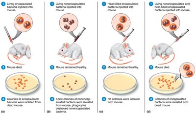
Capsules are a major virulence factor for some bacteria, such as Streptococcus pneumoniae. The classic Griffith experiment demonstrated that non-virulent bacteria could acquire virulence by uptake of genetic material from heat-killed virulent bacteria, highlighting the importance of the capsule in pathogenicity.
Capsule: A polysaccharide layer that impairs phagocytosis and enhances survival in the host.
Transformation: Uptake of genetic material from the environment, leading to new traits such as capsule formation.
Virulence Gene: The gene for capsule synthesis is transferred, enabling non-encapsulated bacteria to become virulent.
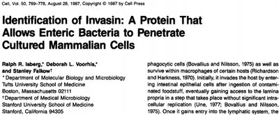
Invasins and Host Cell Penetration
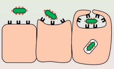
Some bacteria produce invasins, which are proteins that facilitate entry into host cells by rearranging the host cytoskeleton. This process is crucial for pathogens that need to invade tissues or evade immune responses.
Invasins: Surface proteins that induce actin rearrangement and membrane ruffling in host cells.
Example: Yersinia species use invasins to penetrate intestinal epithelial cells.
Intracellular Survival and Evasion of Host Defenses
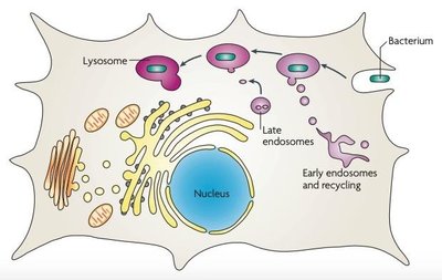
Some pathogens survive within host cells by avoiding lysosomal fusion or escaping from the phagosome. This allows them to evade immune detection and persist within the host.
Prevention of Lysosome Fusion: Bacteria such as Salmonella enterica and Legionella pneumophila prevent fusion of the phagosome with lysosomes, creating a protected niche for replication.
Escape from Phagosome: Shigella flexneri and Listeria monocytogenes escape into the cytoplasm before lysosomal fusion.
Benefits: Access to host nutrients, protection from immune cells, and avoidance of antimicrobial molecules.
Acquisition of Host Nutrients: Siderophores
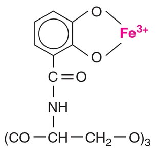
Iron is essential for bacterial growth, but is limited in the host. Pathogens secrete siderophores, which are high-affinity iron-chelating compounds that scavenge iron from host proteins.
Siderophores: Molecules that bind iron more tightly than host proteins, facilitating iron uptake by bacteria.
Example: Enterobactin produced by Escherichia coli and Salmonella enterica.
Bacterial Toxins: Exotoxins and Endotoxins
Bacterial toxins are major virulence factors that damage host tissues and disrupt normal physiological processes. Exotoxins are secreted proteins, while endotoxins are components of the outer membrane of Gram-negative bacteria.
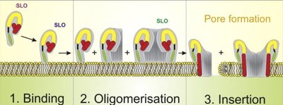
Exotoxins: Proteins secreted by bacteria; include A-B toxins, membrane-disrupting toxins, and superantigens.
Endotoxins: Lipid A component of lipopolysaccharide (LPS) in Gram-negative bacteria; released upon cell lysis.
Effects: Fever, tissue damage, shock, and immune system activation.

Innate Immunity: Nonspecific Defenses of the Host
Physical and Chemical Barriers
Innate immunity provides the first line of defense against pathogens through physical and chemical barriers. These mechanisms are present at birth and act rapidly to prevent infection.
Skin: Composed of the dermis and epidermis; keratinized cells form a tough barrier.
Mucous Membranes: Line body tracts and secrete mucus to trap microbes.
Ciliary Escalator: Moves mucus and trapped microbes out of the respiratory tract.
Chemical Factors: Sebum, lysozyme, low pH of gastric acid and vaginal secretions inhibit microbial growth.
Cellular Components of Innate Immunity
Blood contains various cells involved in innate immunity, including granulocytes and agranulocytes. These cells perform functions such as phagocytosis, inflammation, and coordination of immune responses.
Granulocytes: Neutrophils (phagocytic), basophils (release histamine), eosinophils (against parasites), mast cells (inflammatory mediators).
Agranulocytes: Monocytes (mature into macrophages), dendritic cells (antigen presentation), lymphocytes (T cells, B cells, NK cells).
Phagocytosis and Inflammation
Phagocytosis is the process by which phagocytes ingest and destroy microbes. Inflammation is a local response to infection or injury, characterized by redness, heat, swelling, pain, and loss of function.
Phagocytosis Steps: Chemotaxis, adherence, ingestion (opsonization), digestion (phagolysosome), exocytosis.
Inflammation: Involves vasodilation, increased permeability, and recruitment of immune cells.
Complement System and Antimicrobial Proteins
The complement system consists of serum proteins that enhance immune responses through a cascade of activation. Antimicrobial peptides and iron-binding proteins also contribute to innate defense.
Complement Pathways: Classical (antibody-mediated), alternative (direct activation), lectin (mannose-binding lectin).
Antimicrobial Peptides: Defensins, cathelicidins disrupt microbial membranes.
Iron-Binding Proteins: Transferrin, lactoferrin, ferritin, hemoglobin limit iron availability to microbes.
Interferons
Interferons are cytokines produced in response to viral infections. They induce the production of antiviral proteins in neighboring cells, inhibiting viral replication.
IFN-α and IFN-β: Induce antiviral states in cells.
IFN-γ: Activates macrophages and neutrophils.
