 Back
BackMicrobial Mechanisms of Pathogenicity: Bacterial Pathogenesis and Host Defense Evasion
Study Guide - Smart Notes
 Tailored notes based on your materials, expanded with key definitions, examples, and context.
Bacterial Pathogenesis
Overview of Pathogenesis
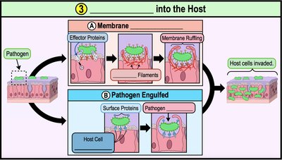
Bacterial pathogenesis refers to the process by which bacteria cause disease in a host. This process involves several distinct steps, each critical for the establishment and progression of infection.
Step 1: Exposure to Pathogen – The host encounters the pathogen, often through a portal of entry.
Step 2: Adhesion to Host – The pathogen adheres to host tissues, typically via specific molecules called adhesins.
Step 3: Invasion into Host – The pathogen invades host cells or tissues, often using mechanisms like induced endocytosis.
Step 4: Multiplication Inside Host – The pathogen multiplies within the host, causing local, focal, or systemic infections.
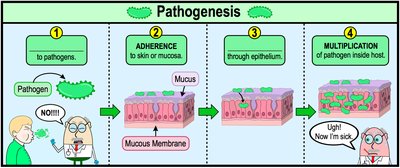
Step 1: Exposure to Pathogen
The initial step in pathogenesis is exposure to the pathogen. Not all exposures result in infection; the pathogen must enter the host through a portal of entry, such as the skin, mucous membranes, or parenteral routes.
Portal of Entry: Sites where host tissues contact the external environment (e.g., respiratory tract, digestive tract).
Example: The respiratory tract is a common portal of entry for pathogens like SARS-CoV-2.
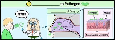
Step 2: Adhesion to Host
After entering the host, pathogens must adhere to host cells to establish infection. Adhesion is mediated by adhesins, which are molecules on the pathogen's surface that bind to specific receptors on host cells.
Adhesins: Found on bacterial pili, fimbriae, and flagella; bind to host cell receptors.
Specificity: Adhesin-receptor binding determines which cell types a pathogen can infect.
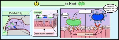
Step 3: Invasion into Host Cell or Tissues
Once adhered, pathogens invade host cells or tissues. This can occur via induced endocytosis, where the pathogen manipulates the host cell to engulf it, or by expressing surface proteins that facilitate entry.
Induced Endocytosis: Pathogen secretes effector proteins, causing membrane ruffling and engulfment.
Membrane Ruffling: Rearrangement of actin filaments in the host cell, leading to internalization of the pathogen.
Surface Proteins: Pathogen binds to host cell receptors and is engulfed.
Step 4: Multiplication Inside Host
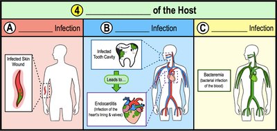
After invasion, pathogens multiply within the host, causing infection. The infection can be classified as local, focal, or systemic based on its spread.
Local Infection: Confined to a specific area near the portal of entry.
Focal Infection: Localized infection spreads to another location, often via the bloodstream.
Systemic Infection: Infection disseminates throughout the body.
Host Defense Evasion Mechanisms
Overview of Host Defenses
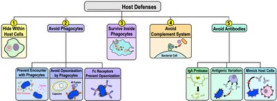
Pathogens have evolved various mechanisms to evade host immune defenses, allowing them to survive and proliferate within the host.
Hide Within Host Cells
Avoid Phagocytosis
Survive Inside Phagocytes
Avoid Complement System
Avoid Antibodies
Hiding Within Host Cells
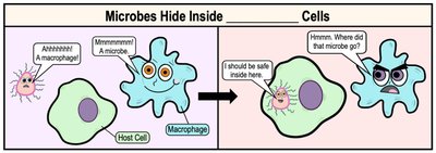
Some pathogens evade immune detection by hiding within host cells, protecting themselves from antibodies and phagocytic cells.
Intracellular Pathogenesis: Pathogens establish infection within host cells, avoiding immune surveillance.
Advantages: Avoids antibodies, opsonization proteins, complement system, and allows dormancy.
Avoiding Phagocytosis
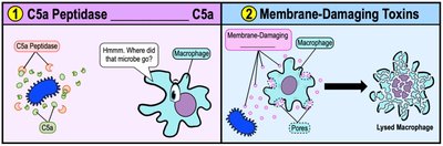
Pathogens can avoid being engulfed and destroyed by phagocytic cells through several strategies.
C5a Peptidase: Degrades C5a, a chemoattractant for phagocytes, reducing immune cell recruitment.
Membrane-Damaging Toxins: Kill phagocytes by forming pores or damaging membranes.
Preventing Opsonization
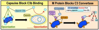
Opsonization enhances phagocytosis by marking pathogens for destruction. Some bacteria prevent opsonization by producing capsules or M proteins.
Capsules: Prevent C3b binding, making it harder for phagocytes to recognize and engulf pathogens.
M Protein: Binds regulatory proteins that degrade C3 convertase, inhibiting C3b production.
Fc Receptors and Opsonization
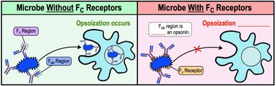
Some pathogens express Fc receptors that bind the Fc region of antibodies, preventing them from acting as opsonins and interfering with phagocytosis.
Fc Receptors: Orient antibodies so the Fab region points outward, which does not promote opsonization.
Surviving Inside Phagocytic Cells
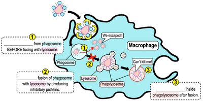
After being engulfed by phagocytes, some bacteria survive by escaping the phagosome, preventing fusion with lysosomes, or resisting digestive enzymes.
Escape from Phagosome: Pathogen forms pores and escapes before lysosome fusion.
Prevent Fusion: Produces proteins that block phagosome-lysosome fusion.
Resist Enzymes: Withstands hydrolytic enzymes inside the phagolysosome.
Avoiding the Complement System
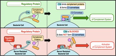
The complement system can cause inflammation, opsonization, or formation of membrane-attack complexes (MAC). Serum-resistant bacteria avoid complement activation by binding and inhibiting C3b.
Regulatory Proteins: Bind and inhibit C3b, preventing C3 convertase formation and complement activation.
Avoiding Antibodies
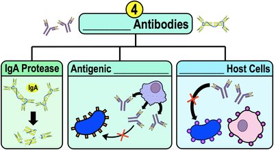
Bacteria have evolved mechanisms to avoid being tagged by antibodies, including production of IgA proteases, antigenic variation, and mimicking host molecules.
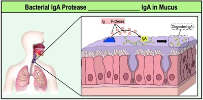
IgA Protease: Degrades IgA antibodies in mucous membranes, reducing immune response.
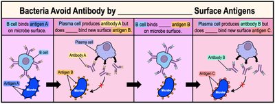
Antigenic Variation: Alters surface antigens to evade antibody recognition.
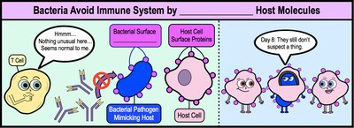
Mimicry: Produces antigens similar to host molecules, avoiding immune detection.
IgA Protease Mechanism
IgA protease specifically targets and degrades IgA antibodies found in mucous membranes, allowing pathogens to evade local immune defenses.
Antigenic Variation
Antigenic variation allows pathogens to change their surface antigens, staying ahead of the host's adaptive immune response. This is why vaccines for certain diseases, like influenza, must be updated regularly.
Mimicking Host Molecules
Some bacteria mimic host cell surface proteins, making it difficult for the immune system to distinguish them from self-cells due to immune tolerance mechanisms.
Pathogenic Toxins
Overview of Toxins
Pathogens produce toxins that damage host tissues and facilitate infection. Toxins are classified as exotoxins or endotoxins.
Exotoxins: Soluble proteins released by pathogens; highly specific in their action.
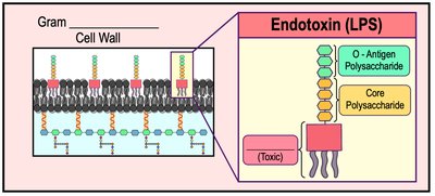
Endotoxins: Lipopolysaccharide (LPS) found in the outer membrane of gram-negative bacteria; triggers systemic responses.
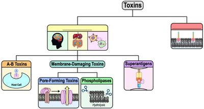
Exotoxins: Types and Effects
Exotoxins are grouped based on the tissue they affect:
Neurotoxins: Affect the nervous system, causing paralysis.
Enterotoxins: Affect the digestive tract, causing diarrhea and vomiting.
Cytotoxins: Affect various cell types, causing cell death or dysfunction.
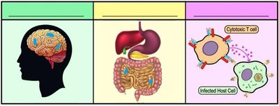
A-B Toxins
A-B toxins consist of two subunits: the A (active) subunit, which is enzymatic and causes damage, and the B (binding) subunit, which determines cell specificity. The process involves binding, endocytosis, and cellular targeting.
B Subunit: Binds to host cell receptor.
A Subunit: Enters cell and disrupts cellular functions.
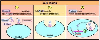
Membrane-Damaging Toxins
These exotoxins disrupt the cytoplasmic membrane, causing cell lysis. They include pore-forming toxins and phospholipases.
Pore-Forming Toxins: Create pores in the membrane, leading to cell death.
Phospholipases: Hydrolyze phospholipids, disrupting membrane integrity.
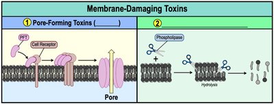
Superantigens
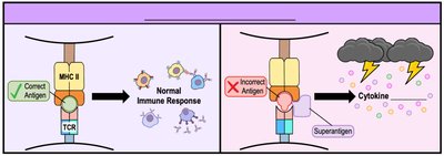
Superantigens are exotoxins that overstimulate T helper cells, causing excessive cytokine release (cytokine storm), which can be life-threatening.
Mechanism: Bind MHC II on antigen-presenting cells, causing inappropriate T cell activation.
Effects: Fever, inflammation, shock.
Endotoxins
Endotoxins are lipopolysaccharides (LPS) found in the outer membrane of gram-negative bacteria. The toxic component, Lipid A, triggers inflammatory responses and can cause septic shock.
Lipid A: Released during bacterial multiplication or lysis; causes fever, anemia, inflammation, and cytokine storm.
Septic Shock: Systemic response affecting multiple organs.
Exotoxins vs. Endotoxins
Comparison Table
Exotoxins and endotoxins differ in their structure, source, toxicity, and effects.
Feature | Exotoxin | Endotoxin |
|---|---|---|
Source | Gram-positive and Gram-negative bacteria | Gram-negative bacteria |
Structure | Protein | Lipopolysaccharide (LPS) |
Toxicity | Highly toxic, lethal in small doses | Less toxic, lethal in large doses |
Heat Stability | Heat labile | Heat stable |
Effects | Specific (e.g., neurotoxins, enterotoxins) | General (fever, shock) |
*Additional info: Table inferred from textbook context and practice questions.*
Viruses Evading the Immune Response
Mechanisms of Viral Evasion
Viruses employ several strategies to evade host immune defenses, ensuring successful replication and persistence.
Prevent Antiviral Effects: Coat viral RNA to avoid detection by pattern recognition receptors (PRRs); inhibit apoptosis by suppressing host gene expression.
Interfere with Antigen Presentation: Block movement of MHC I molecules to cell surface; produce "fake" MHC I to trick NK cells.
Avoid Antibodies: Rapid mutation and antigenic variation; antibody-dependent enhancement (ADE) allows viruses to use antibodies to enter macrophages and replicate.
*Additional info: Viral evasion mechanisms are analogous to bacterial strategies, including intracellular survival and antigenic variation.*
