Back
Microbial Mechanisms of Pathogenicity: How Microbes Cause Disease
Study Guide - Smart Notes
 Tailored notes based on your materials, expanded with key definitions, examples, and context.
Microbial Mechanisms of Pathogenicity
Introduction to Pathogenicity and Virulence
Pathogenicity refers to a microorganism's ability to cause disease by overcoming host defenses, while virulence describes the degree of pathogenicity. For a microbe to cause disease, it must enter the host, adhere to host tissues, penetrate or evade host defenses, and damage host tissues. Some microbes cause disease not by direct damage, but by the accumulation of their waste products.
Portals of Entry
Main Portals of Entry
Pathogens enter the human body through specific routes known as portals of entry. The main portals include mucous membranes, skin, and the parenteral route (direct deposition into tissues when barriers are penetrated). Most pathogens have a preferred portal of entry, which is essential for their ability to cause disease.
Mucous membranes: Respiratory, gastrointestinal, genitourinary tracts, and conjunctiva.
Skin: Usually impenetrable unless there are openings.
Parenteral route: Through punctures, injections, bites, cuts, wounds, or surgery.
Respiratory Tract
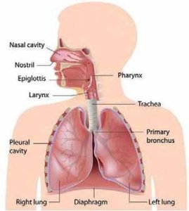
The respiratory tract is the easiest and most common portal of entry for many pathogens, which are inhaled into the nose or mouth in droplets of moisture. Diseases include the common cold, pneumonia, tuberculosis, influenza, and measles.
Gastrointestinal Tract
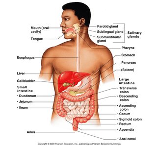
Pathogens enter via food, water, or contaminated fingers. Most are destroyed by stomach acid, but those that survive can cause diseases such as polio, hepatitis A, and typhoid fever. These pathogens are often eliminated with feces and can be transmitted to others via contaminated water, food, or fingers.
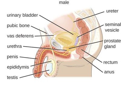
Genitourinary Tract
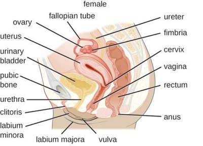
This portal is associated with sexually transmitted infections (STIs). Some microbes penetrate unbroken mucous membranes, while others require a cut or abrasion. Examples include HIV, genital warts, chlamydia, herpesvirus-2, syphilis, and gonorrhea.
Skin
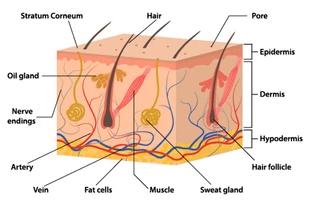
The skin is an important defense against disease. Unbroken skin is generally impenetrable, but some microbes can enter through hair follicles, sweat gland ducts, or by boring through intact skin (e.g., hookworm larvae). Some fungi infect the skin or grow on keratin.
Parenteral Route
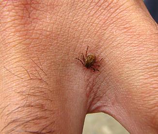
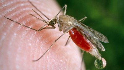
Microorganisms can be deposited directly into tissues via punctures, injections, bites, cuts, wounds, or surgery. Diseases such as HIV, hepatitis, tetanus, and gangrene can be transmitted this way.
Preferred Portal of Entry
Pathogens often require a specific portal of entry to cause disease. Entry through a different portal may not result in disease.
Numbers of Invading Microbes
ID50 and LD50
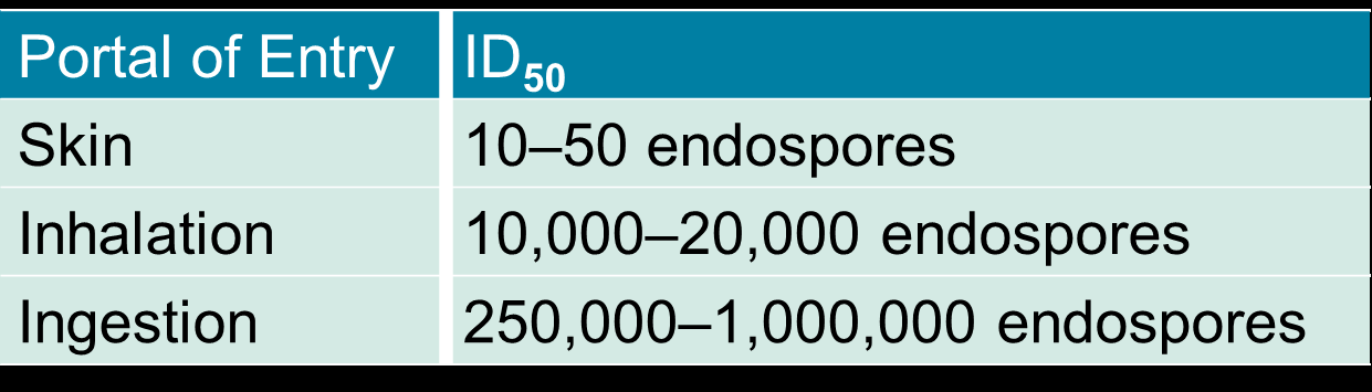
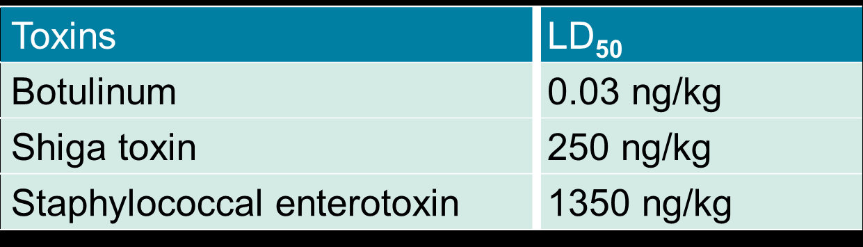
The likelihood of disease increases with the number of invading pathogens. Two important measures are:
ID50: Infectious dose for 50% of a sample population (measures virulence).
LD50: Lethal dose for 50% of a sample population (measures toxin potency).

Adherence to Host Tissues
Mechanisms of Adherence
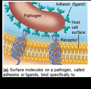
Most pathogens must attach to host tissues to establish infection. This process, called adherence (or adhesion), involves adhesins (ligands) on the pathogen binding to specific receptors on host cells. Structures involved include the glycocalyx and fimbriae. Microbes may also form biofilms, which are communities that share nutrients and enhance survival.
Penetration or Evasion of Host Defenses
Virulence Factors
Capsules: Glycocalyx layers that impair phagocytosis.
Cell wall components: M protein (resists phagocytosis), Opa protein (attachment), mycolic acid (resists digestion).
Enzymes: Exoenzymes such as coagulases, kinases, hyaluronidase, collagenase, and IgA proteases aid in invasion and evasion.
Antigenic variation: Pathogens alter their surface antigens to evade immune detection.
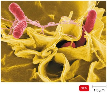
Invasins: Surface proteins that rearrange host cell actin, causing membrane ruffling and facilitating entry (e.g., Shigella, Listeria).
Biofilms: Protect bacteria from phagocytosis and enhance resistance.
How Bacterial Pathogens Damage Host Cells
Mechanisms of Damage
Once inside the host, pathogens can damage cells in several ways:
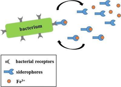
Using the host’s nutrients (e.g., iron via siderophores).
Causing direct damage at the site of invasion.
Producing toxins that affect distant sites.
Inducing hypersensitivity reactions.
Using Host Nutrients: Siderophores
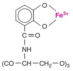
Iron is essential for most pathogenic bacteria. Siderophores are proteins secreted by pathogens that bind iron more tightly than host cells, allowing bacteria to acquire this vital nutrient.
Direct Damage
Pathogens can disrupt host cell function, use host nutrients, produce waste products, and multiply within host cells, causing cell rupture.
Production of Toxins
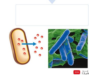
Toxins are poisonous substances produced by microorganisms. They can cause fever, cardiovascular problems, diarrhea, and shock. Toxigenicity is the ability to produce toxins, while toxemia is the presence of toxins in the blood. There are two main types of toxins:
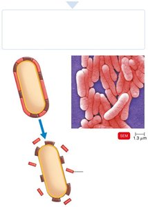
Exotoxins: Proteins secreted by bacteria (mainly Gram-positive) during growth. They are highly toxic and can act repeatedly.
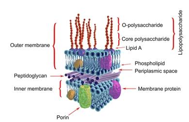
Endotoxins: Lipid A component of lipopolysaccharides (LPS) in the outer membrane of Gram-negative bacteria, released upon cell death.
Exotoxins vs. Endotoxins
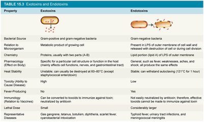
Property | Exotoxins | Endotoxins |
|---|---|---|
Bacterial Source | Gram-positive and Gram-negative bacteria | Gram-negative bacteria |
Relation to Microorganism | Metabolic product of growing cell | Present in LPS of outer membrane; released on cell lysis |
Chemistry | Protein, usually with two parts (A-B) | Lipid portion (Lipid A) of LPS |
Pharmacology (Effect on Body) | Specific for a particular cell function | General, such as fever, weakness, aches, shock |
Heat Stability | Unstable (destroyed at 60–80°C) | Stable (withstands autoclaving) |
Toxicity | High | Low |
Fever-Producing | No | Yes |
Immunogenicity | Can be converted to toxoids for vaccines | Not easily neutralized by antitoxin; no effective toxoid |
Representative Diseases | Gas gangrene, tetanus, botulism, diphtheria, scarlet fever | Typhoid fever, urinary tract infections, meningococcal meningitis |
Endotoxins and the Pyrogenic Response
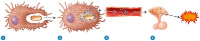
Endotoxins stimulate macrophages to release cytokines, such as interleukin-1 (IL-1) and tumor necrosis factor alpha (TNF-α), which travel to the hypothalamus and induce fever by triggering prostaglandin production.
Portals of Exit
How Pathogens Leave the Host
Pathogens exit the host through specific portals, often the same as their entry points. These include:
Respiratory tract: Coughing and sneezing
Gastrointestinal tract: Feces and saliva
Genitourinary tract: Urine and genital secretions
Skin
Blood: Via arthropod bites or needles
Key Concepts and Summary
Several factors are required for a microbe to cause disease: entry, adherence, penetration/evasion, and damage to host tissues.
Pathogens usually leave the body via specific portals of exit, which are generally the same as their portals of entry.
Understanding microbial pathogenicity is fundamental to understanding infectious disease processes.