 Back
BackMicrobial Mechanisms of Pathogenicity: Study Notes
Study Guide - Smart Notes
 Tailored notes based on your materials, expanded with key definitions, examples, and context.
Microbial Mechanisms of Pathogenicity
Introduction
This chapter explores how microorganisms cause disease, focusing on the mechanisms by which pathogens enter, survive, and damage the host. Understanding these processes is essential for microbiology students to grasp the basis of infectious diseases and host-pathogen interactions.
How Microorganisms Enter a Host
Portals of Entry
Pathogens must enter the host through specific portals to initiate infection. The main portals of entry include:
Mucous membranes: Entry via respiratory, gastrointestinal, and genitourinary tracts. The respiratory tract is the most common portal (e.g., influenza, pneumonia).
Skin: Although unbroken skin is a strong barrier, some microbes enter through hair follicles or sweat gland ducts (e.g., trachoma, conjunctivitis).
Parenteral route: Microbes are deposited directly into tissues beneath the skin or mucous membranes, often via bites, cuts, or injections (e.g., tetanus, hepatitis viruses).
Preferred Portal of Entry
Most pathogens have a preferred portal of entry. If they enter by another route, disease may not occur.
Numbers of Invading Microbes
ID50 and LD50
ID50 (Infectious Dose 50): The number of pathogen cells or virions required to cause infection in 50% of a test population. It measures the virulence of a microbe.
LD50 (Lethal Dose 50): The amount of toxin required to kill 50% of a test population. It measures the potency of a toxin.
Example: The LD50 for botulinum toxin in mice is 0.03 ng/kg, indicating extreme potency.
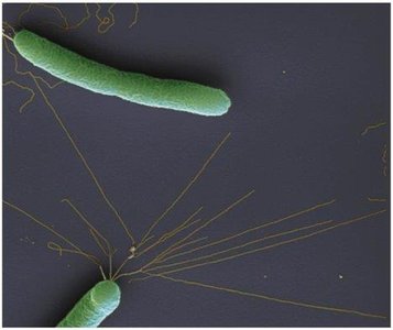
Adherence to Host Tissues
Mechanisms of Adherence
Adherence is a critical step in infection. Pathogens use surface molecules called adhesins (ligands) to bind specifically to complementary receptors on host cells. These adhesins may be located on the glycocalyx or fimbriae.
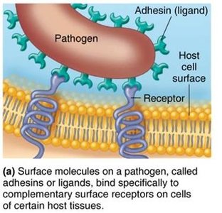
If adhesins or receptors are altered, infection can often be prevented.
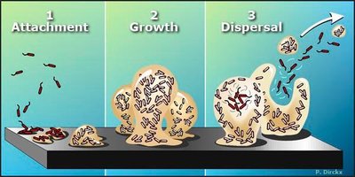
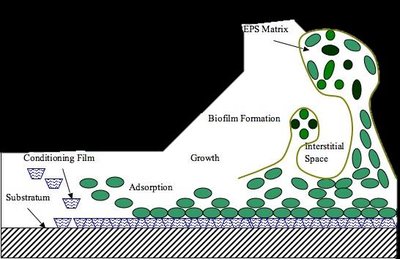
Microbes can form biofilms, communities that adhere to surfaces and are protected by a glycocalyx. Biofilms enhance microbial survival and resistance (e.g., dental plaque).
How Pathogens Penetrate Host Defenses
Capsules and Cell Wall Components
Capsules: Glycocalyx layers that impair phagocytosis (e.g., Streptococcus pneumoniae).
M protein: Resists phagocytosis (e.g., Streptococcus pyogenes).
Opa protein: Facilitates attachment to host cells (e.g., Neisseria gonorrhoeae).
Mycolic acid: Waxy lipid in cell walls resists digestion (e.g., Mycobacterium tuberculosis).
Enzymes as Virulence Factors
Coagulases: Clot fibrinogen in blood, protecting bacteria from immune cells.
Kinases: Digest fibrin clots, allowing spread of infection.
Hyaluronidase: Digests polysaccharides holding cells together, facilitating tissue invasion.
Collagenase: Breaks down collagen in connective tissue.
IgA proteases: Destroy IgA antibodies, aiding in immune evasion.
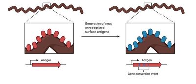
Antigenic Variation
Some pathogens alter their surface antigens, rendering antibodies ineffective. This allows them to evade the immune response.
Penetration into Host Cells
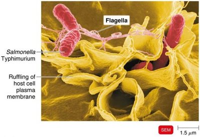
Invasins: Surface proteins that rearrange actin filaments, causing membrane ruffling and facilitating entry (e.g., Salmonella).
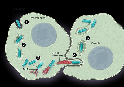
Some bacteria use actin to move between cells (e.g., Shigella, Listeria).
Biofilms and Immune Evasion
Biofilms protect bacteria from phagocytosis and immune responses due to their extracellular polymeric substance (EPS) matrix.
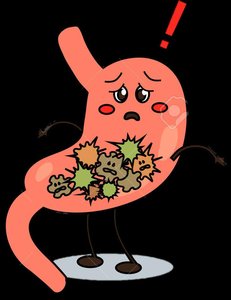
How Bacterial Pathogens Damage Host Cells
Mechanisms of Host Cell Damage
Using host nutrients (e.g., iron via siderophores)
Direct damage to host cells
Production of toxins
Inducing hypersensitive reactions
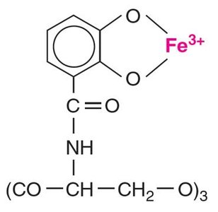
Siderophores
Siderophores are proteins secreted by pathogens to scavenge iron from the host, which is essential for bacterial growth.
Toxins
Toxins: Poisonous substances produced by microorganisms that can cause fever, shock, and other symptoms.
Toxigenicity: The ability to produce toxins.
Toxemia: Presence of toxins in the blood.
Intoxications: Disease caused by toxins without microbial growth.
Exotoxins
Proteins secreted by bacteria, usually Gram-positive, that are highly potent and specific in action.
Genes for exotoxins are often carried on plasmids.
Types of exotoxins:
A-B toxins: Consist of an active (A) and binding (B) component (e.g., diphtheria toxin).
Membrane-disrupting toxins: Cause cell lysis (e.g., hemolysins, leukocidins).
Superantigens: Trigger excessive immune responses, leading to shock.
Genotoxins: Damage DNA, potentially leading to cancer.
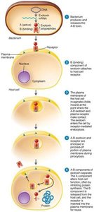
Table: Diseases Caused by Exotoxins
Disease | Bacterium | Type of Exotoxin | Mechanism |
|---|---|---|---|
Botulism | Clostridium botulinum | A-B | Neurotoxin prevents nerve impulse transmission; flaccid paralysis. |
Tetanus | Clostridium tetani | A-B | Blocks muscle relaxation pathway; uncontrollable contractions. |
Diphtheria | Corynebacterium diphtheriae | A-B | Inhibits protein synthesis in nerve, heart, kidney cells. |
Toxic shock syndrome | Staphylococcus aureus | Superantigen | Causes fluid loss, low blood pressure. |
Traveler’s diarrhea | E. coli, Shigella spp. | A-B | Enterotoxin causes diarrhea. |
Anthrax | Bacillus anthracis | A-B | Shock, reduced immune response. |
Endotoxins
Lipid A component of lipopolysaccharide (LPS) in Gram-negative bacteria.
Released during bacterial death or multiplication.
Cause general symptoms: fever, shock, weakness, and can trigger miscarriages.
Detected by the Limulus amebocyte lysate (LAL) assay.
Table: Comparison of Exotoxins and Endotoxins
Property | Exotoxins | Endotoxins |
|---|---|---|
Chemistry | Proteins (A-B structure) | Lipid A (LPS) |
Source | Gram-positive & Gram-negative | Gram-negative |
Heat Stability | Unstable (destroyed at 60–80°C) | Stable (withstands autoclaving) |
Toxicity | High | Low |
Fever Producing | No | Yes |
Immunology | Can be neutralized by antitoxin | Not easily neutralized |
Lethal Dose | Small | Larger |
Pathogenic Properties of Viruses, Fungi, Protozoa, Helminths, and Algae
Viruses
Cause cytopathic effects (CPE) such as cell death, inclusion bodies, syncytia formation, and chromosomal changes.
Alpha and beta interferons produced by infected cells protect neighboring cells by inhibiting viral protein synthesis and inducing apoptosis.
Fungi
Produce toxic metabolic products (e.g., aflatoxin, ergot alkaloids).
Can provoke allergic responses and inhibit protein synthesis (e.g., trichothecene toxins).
Capsules prevent phagocytosis.
Protozoa
Cause disease by growing in host tissues, producing waste products, and evading immune responses via antigenic variation.
Helminths
Use host tissues for growth, produce large masses, and release waste products that cause symptoms.
Algae
Some produce neurotoxins (e.g., saxitoxin) that cause paralytic shellfish poisoning.
Portals of Exit
Major Portals of Exit
Pathogens leave the host via specific portals, often the same as the portals of entry:
Respiratory tract: Coughing and sneezing
Gastrointestinal tract: Feces and saliva
Genitourinary tract: Urine and genital secretions
Skin
Blood: Via arthropod bites or contaminated needles
Additional info: This guide expands on the original notes with definitions, examples, and tables for clarity and exam preparation.
