 Back
Back15 Microbial Mechanisms of Pathogenicity: Study Notes
Study Guide - Smart Notes
 Tailored notes based on your materials, expanded with key definitions, examples, and context.
Microbial Mechanisms of Pathogenicity
Introduction
This chapter explores how microorganisms cause disease, focusing on the mechanisms by which pathogens enter, survive, and damage their hosts. Understanding these processes is essential for microbiology students to grasp the basis of infectious diseases and host-pathogen interactions.
How Microorganisms Enter a Host
Portals of Entry
Pathogens must enter the host through specific portals to initiate infection. The main portals of entry include:
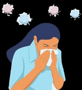
Mucous membranes: Entry via respiratory, gastrointestinal, and genitourinary tracts. The respiratory tract is the most common portal (e.g., influenza, pneumonia).
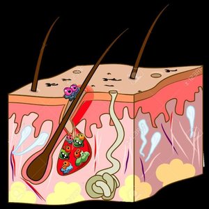
Skin: Although unbroken skin is a strong barrier, some microbes enter through hair follicles or sweat gland ducts (e.g., trachoma, conjunctivitis).
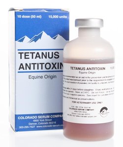
Parenteral route: Microbes are deposited directly into tissues beneath the skin or mucous membranes via bites, cuts, or injections (e.g., tetanus, hepatitis viruses).
ID50 and LD50
The virulence of a microbe is often measured by:
ID50 (Infectious Dose 50): The number of pathogen cells or virions required to cause infection in 50% of a test population.
LD50 (Lethal Dose 50): The amount of toxin required to kill 50% of a test population.
Lower ID50 or LD50 values indicate higher virulence or toxicity.

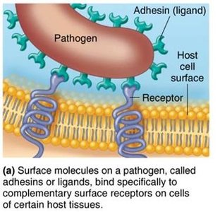
Adherence to Host Tissues
Most pathogens must adhere to host tissues to establish infection. This process involves:
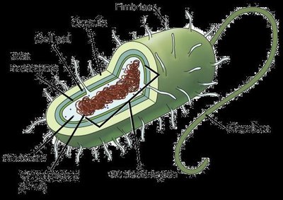
Adhesins (ligands): Surface molecules on pathogens that bind specifically to complementary receptors on host cells. Adhesins are often located on the glycocalyx or fimbriae.
If adherence is blocked, infection can often be prevented.
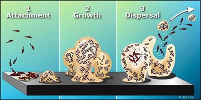
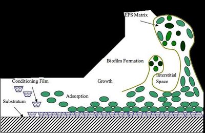
Biofilms
Microbes can form biofilms, which are communities of microorganisms attached to a surface and encased in a self-produced matrix. Biofilms enhance adherence and protect microbes from host defenses.
Examples: Dental plaque, algae on swimming pool walls.
How Pathogens Penetrate Host Defenses
Capsules and Cell Wall Components
Some bacteria evade host defenses using:
Capsules: Glycocalyx layers that impair phagocytosis (e.g., Streptococcus pneumoniae).
Cell wall components:
M protein: Resists phagocytosis (Streptococcus pyogenes).
Opa protein: Aids attachment (Neisseria gonorrhoeae).
Mycolic acid: Waxy lipid resists digestion (Mycobacterium tuberculosis).
Enzymes as Virulence Factors
Bacteria may secrete enzymes that facilitate invasion and damage:
Coagulases: Clot fibrinogen in blood.
Kinases: Digest fibrin clots.
Hyaluronidase: Digests polysaccharides holding cells together.
Collagenase: Breaks down collagen in connective tissue.
IgA proteases: Destroy IgA antibodies.
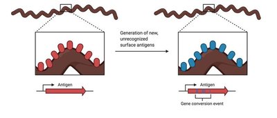
Antigenic Variation
Some pathogens evade the immune system by altering their surface antigens, rendering antibodies ineffective. This process is called antigenic variation.
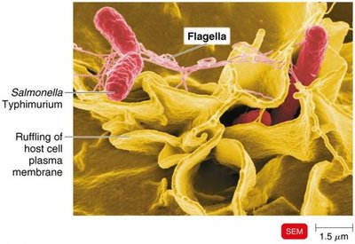
Penetration into Host Cells
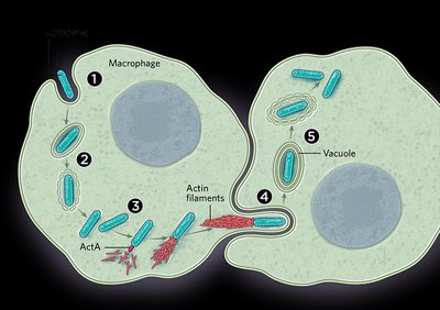
Bacteria can invade host cells by manipulating the host cytoskeleton:
Invasins: Surface proteins that rearrange actin filaments, causing membrane ruffling (e.g., Salmonella entry).
Some bacteria use actin to move between cells (e.g., Shigella, Listeria).
Biofilms and Evasion of Phagocytosis
Biofilms protect bacteria from phagocytosis by shielding antigens and sometimes killing phagocytes with their extracellular polymeric substance (EPS).
How Bacterial Pathogens Damage Host Cells
Mechanisms of Host Cell Damage
Pathogens damage host cells by:
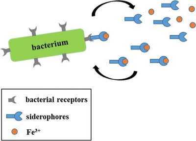
Using host nutrients (e.g., iron via siderophores)
Direct damage (disrupting cell function, producing waste, causing cell rupture)
Producing toxins (exotoxins and endotoxins)
Inducing hypersensitive reactions
Siderophores
Siderophores are proteins secreted by pathogens to scavenge iron from the host, which is essential for bacterial growth.

Toxins
Toxins are poisonous substances produced by microbes that can cause fever, shock, diarrhea, and other symptoms. The ability to produce toxins is called toxigenicity, and the presence of toxins in blood is toxemia.
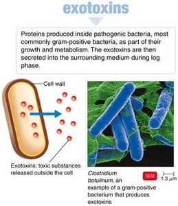
Exotoxins
Exotoxins are proteins secreted by bacteria (mainly Gram-positive) that are highly potent and specific in their action. Genes for exotoxins are often carried on plasmids.
Antitoxins: Antibodies that neutralize exotoxins.
Toxoids: Inactivated exotoxins used in vaccines.
Types of Exotoxins
A-B toxins: Consist of an active (A) and binding (B) component. Example: diphtheria toxin.
Membrane-disrupting toxins: Cause cell lysis by disrupting plasma membranes (e.g., hemolysins, leukocidins).
Superantigens: Trigger excessive immune responses, leading to fever, shock, and sometimes death.
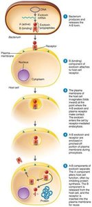
Table: Diseases Caused by Exotoxins
Disease | Bacterium | Type of Exotoxin | Mechanism |
|---|---|---|---|
Botulism | Clostridium botulinum | A-B | Neurotoxin prevents nerve impulse transmission; flaccid paralysis. |
Tetanus | Clostridium tetani | A-B | Blocks muscle relaxation pathway; uncontrollable contractions. |
Diphtheria | Corynebacterium diphtheriae | A-B | Inhibits protein synthesis in nerve, heart, kidney cells. |
Toxic shock syndrome | Staphylococcus aureus | Superantigen | Causes fluid loss, low blood pressure. |
Traveler’s diarrhea | E. coli, Shigella spp. | A-B | Enterotoxin causes diarrhea. |
Endotoxins
Endotoxins are lipid components (Lipid A) of the outer membrane of Gram-negative bacteria. They are released when bacteria die and can cause fever, shock, and other systemic effects.
All endotoxins produce similar symptoms regardless of bacterial species.
Endotoxins are heat-stable and not easily neutralized by antitoxins.
Table: Comparison of Exotoxins and Endotoxins
Property | Exotoxins | Endotoxins |
|---|---|---|
Chemistry | Proteins (A-B structure) | Lipid A (LPS) |
Source | Gram-positive & Gram-negative | Gram-negative only |
Heat Stability | Unstable (destroyed at 60–80°C) | Stable (withstands 121°C) |
Toxicity | High | Low |
Fever-producing | No | Yes |
Immunology | Can be neutralized by antitoxin | Not easily neutralized |
Lethal Dose | Small | Large |
Plasmids, Lysogeny, and Pathogenicity
Plasmids may carry genes for toxins, antibiotic resistance, and enzymes. Lysogenic conversion (integration of a prophage) can turn non-pathogenic bacteria into pathogens by introducing new virulence factors.
Pathogenic Properties of Viruses, Fungi, Protozoa, Helminths, and Algae
Pathogenic Properties of Viruses
Viruses cause disease through cytopathic effects (CPE), which are visible changes in host cells due to viral infection. Examples include:
Stopping cell synthesis
Causing lysosome release
Forming inclusion bodies
Fusing cells to form syncytia
Inducing chromosomal changes
Loss of contact inhibition (leading to cancer)
Alpha and beta interferons produced by infected cells protect neighboring cells by inhibiting viral protein synthesis and inducing apoptosis in infected cells.
Pathogenic Properties of Fungi
Fungi may cause disease by:
Producing toxic metabolic products (e.g., trichothecene toxins)
Provoking allergic responses
Producing proteases that modify host membranes
Forming capsules to evade phagocytosis
Producing mycotoxins (e.g., aflatoxin, ergot alkaloids, phalloidin, amanitin)
Pathogenic Properties of Protozoa
Protozoa cause symptoms by:
Digesting host cells and tissues
Growing inside phagocytes
Undergoing antigenic variation
Pathogenic Properties of Helminths
Helminths (parasitic worms) cause disease by:
Using host tissues for growth
Producing large masses that cause cellular damage
Releasing waste products that provoke symptoms
Pathogenic Properties of Algae
Some algae produce neurotoxins, such as saxitoxin, which can cause paralytic shellfish poisoning (PSP). These toxins are heat-stable and not destroyed by cooking.
Portals of Exit
Major Portals of Exit
Pathogens leave the host through specific portals, which often mirror the portals of entry:
Respiratory tract: Coughing and sneezing
Gastrointestinal tract: Feces and saliva
Genitourinary tract: Urine and genital secretions
Skin
Blood: Via arthropod bites or contaminated needles
Additional info: This guide expands on the original notes with definitions, examples, and tables for clarity and exam preparation.
