Back
BackMicrobial Mechanisms of Pathogenicity: Study Notes
Study Guide - Smart Notes
 Tailored notes based on your materials, expanded with key definitions, examples, and context.
Microbial Mechanisms of Pathogenicity
Introduction
This chapter explores how microorganisms cause disease, focusing on the mechanisms by which pathogens enter, survive, and damage the host. Understanding these processes is essential for microbiology students to grasp the basis of infectious diseases and host-pathogen interactions.
How Microorganisms Enter a Host
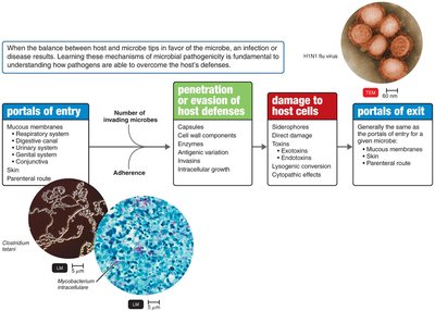
Portals of Entry
Pathogens must enter the host through specific portals to initiate infection. The main portals of entry include:
Mucous membranes: Respiratory tract (most common), digestive canal, genitourinary system, and conjunctiva.
Skin: Usually impenetrable unless through hair follicles or sweat gland ducts.
Parenteral route: Direct deposition into tissues beneath the skin or mucous membranes via punctures, injections, bites, wounds, or surgery.
Most pathogens have a preferred portal of entry, which is critical for their ability to cause disease.
Numbers of Invading Microbes
ID50 (Infectious Dose 50): The number of microbes required to cause infection in 50% of a sample population. It measures the virulence of a microbe.
LD50 (Lethal Dose 50): The amount of toxin required to kill 50% of a sample population. It measures the potency of a toxin.
Example: Bacillus anthracis has different ID50 values depending on the portal of entry (e.g., 10–50 endospores via skin, 10,000–20,000 via inhalation).
Adherence
Pathogens must attach to host tissues to establish infection. This process, called adherence, involves:
Adhesins (ligands): Surface molecules on pathogens that bind to specific receptors on host cells.
Examples:
Glycocalyx of Streptococcus mutans enables adherence to teeth.
Fimbriae of Actinomyces adhere to the glycocalyx of S. mutans.
Viral spikes (e.g., SARS-CoV-2) bind to ACE2 receptors on host cells.
Penetration or Evasion of Host Defenses
Capsules
Some bacteria produce a glycocalyx capsule around their cell wall, which impairs phagocytosis by host immune cells.
Examples: Streptococcus pneumoniae, Haemophilus influenzae, Bacillus anthracis, Yersinia pestis
Cell Wall Components
M protein: Resists phagocytosis (Streptococcus pyogenes).
Opa protein: Facilitates attachment to host cells (Neisseria gonorrhoeae).
Waxy lipid (mycolic acid): Resists digestion by phagocytes (Mycobacterium tuberculosis).
Enzymes
Coagulases: Coagulate fibrinogen to form clots.
Kinases: Digest fibrin clots.
Hyaluronidase: Digests hyaluronic acid, aiding tissue penetration.
Collagenase: Breaks down collagen.
IgA proteases: Destroy IgA antibodies.
Antigenic Variation
Pathogens can alter their surface antigens, rendering host antibodies ineffective. This allows them to evade the immune response.
Examples: Influenza virus, Neisseria gonorrhoeae, Trypanosoma brucei gambiense
Penetration into the Host Cell
Invasins: Surface proteins that rearrange actin filaments, causing membrane ruffling and bacterial engulfment (e.g., Shigella, Listeria).
Survival inside phagocytes: Some bacteria survive by escaping the phagosome, preventing lysosomal fusion, or requiring low pH in the phagolysosome.
Biofilms
Biofilms protect bacteria from antibiotics, disinfectants, and phagocytosis. The extracellular polymeric substance (EPS) shields bacteria, making infections harder to treat.
How Bacterial Pathogens Damage Host Cells
Using the Host’s Nutrients: Siderophores
Pathogens secrete siderophores to bind iron more tightly than host proteins, depriving host cells of this essential nutrient.
Direct Damage
Disruption of host cell function
Consumption of host nutrients
Production of waste products
Multiplication inside host cells, causing cell rupture
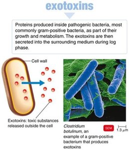
Production of Toxins
Toxins are poisonous substances produced by microorganisms that can cause fever, cardiovascular problems, diarrhea, and shock.
Toxigenicity: Ability to produce toxins
Toxemia: Presence of toxins in the blood
Intoxications: Disease caused by toxins without microbial growth
Exotoxins
Exotoxins are proteins secreted by bacteria, usually highly specific and potent. They can be neutralized by antitoxins and inactivated for use in vaccines (toxoids).
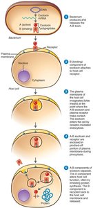
Types of Exotoxins
A-B toxins: Consist of an active (A) and binding (B) component. The B part binds to the host cell, and the A part exerts the toxic effect (e.g., diphtheria toxin).
Genotoxins: Damage DNA, potentially leading to mutations and cancer.
Membrane-disrupting toxins: Lyse host cells by disrupting plasma membranes (e.g., leukocidins, hemolysins, streptolysins).
Superantigens: Cause intense immune responses by stimulating excessive cytokine release, leading to fever, shock, and sometimes death.
Diseases Caused by Exotoxins
Disease | Bacterium | Type of Exotoxin | Mechanism |
|---|---|---|---|
Botulism | Clostridium botulinum | A-B | Neurotoxin; prevents nerve impulse transmission; flaccid paralysis. |
Tetanus | C. tetani | A-B | Neurotoxin; blocks muscle relaxation; uncontrollable contractions. |
Diphtheria | Corynebacterium diphtheriae | A-B | Cytotoxin; inhibits protein synthesis. |
Cholera | Vibrio cholerae | A-B | Enterotoxin; causes diarrhea. |
Anthrax | Bacillus anthracis | A-B | Shock and immune suppression. |
Food poisoning | Staphylococcus aureus | Superantigen | Enterotoxin; diarrhea. |
Toxic shock syndrome | S. aureus | Superantigen | Shock, low blood pressure. |
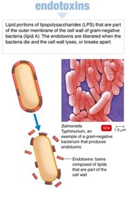
Endotoxins
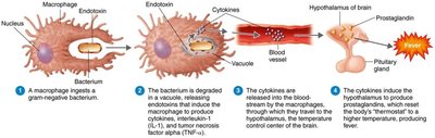
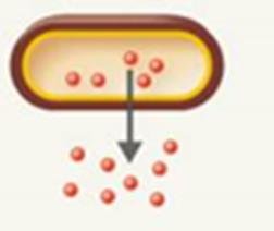
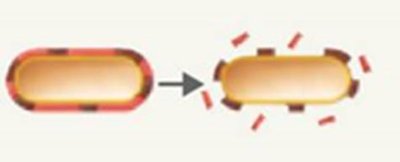
Endotoxins are lipid A components of the lipopolysaccharide (LPS) layer of gram-negative bacteria. They are released when bacteria die and the cell wall breaks apart.
Stimulate macrophages to release cytokines, causing fever, chills, weakness, and shock.
Can cause disseminated intravascular coagulation and endotoxic shock.
May weaken the blood-brain barrier.
Pyrogenic Response
Endotoxins induce fever by stimulating macrophages to release cytokines, which act on the hypothalamus to increase body temperature.
Detection of Endotoxins
The Limulus amebocyte lysate (LAL) assay is used to detect endotoxins. Amebocytes from horseshoe crab blood lyse in the presence of endotoxin, forming a clot.
Comparison of Exotoxins and Endotoxins
Property | Exotoxins | Endotoxins |
|---|---|---|
Bacterial Source | Gram-positive and gram-negative | Gram-negative |
Chemistry | Proteins (A-B structure) | Lipid A of LPS |
Heat Stability | Unstable (destroyed at 60–80°C) | Stable (withstands autoclaving) |
Toxicity | High | Low |
Fever-Producing | No | Yes |
Immunology | Can be neutralized by antitoxin; toxoids for vaccines | Not easily neutralized; no effective toxoids |
Lethal Dose | Small | Large |
Representative Diseases | Tetanus, botulism, diphtheria | Typhoid fever, meningococcal meningitis |
Plasmids, Lysogeny, and Pathogenicity
Plasmids may carry genes for toxins, antibiotic resistance, and virulence factors. Lysogenic conversion can change microbial characteristics, such as toxin production, due to prophage incorporation.
R plasmids: Encode antibiotic resistance.
Virulence plasmids: Encode toxins and enzymes (e.g., tetanus neurotoxin, staphylococcal enterotoxin).
Lysogenic conversion: Can result in new toxin production (e.g., diphtheria toxin).
Pathogenic Properties of Viruses
Mechanisms for Evading Host Defenses
Intracellular location shields viruses from immune detection.
Attachment to host cell surface molecules.
Direct attack on immune system components.
Methylation of viral RNA to mimic host RNA.
Antigenic variation.
Cytopathic Effects (CPE)
Cytopathic effects are visible changes in host cells due to viral infection. They can be cytocidal (cell death) or noncytocidal (cell damage without death).
Disruption of cell junctions
Cytokine storm induction
Inhibition of macromolecular synthesis
Lysosomal enzyme release
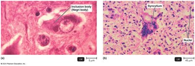
Inclusion body formation (e.g., Negri bodies in rabies)
Syncytium formation (fusion of cells)
Antigenic changes on cell surface
Chromosomal damage
Loss of contact inhibition (unregulated growth, cancer)
Interferon production
Interferons
Alpha and beta interferons are produced by virally infected cells to protect neighboring cells by inhibiting viral protein synthesis and inducing apoptosis. Some viruses can evade interferon effects.
Examples of Cytopathic Effects
Virus (Genus) | Cytopathic Effect |
|---|---|
Poliovirus (Enterovirus C) | Cytocidal (cell death) |
Genital warts virus (Alphapapillomavirus) | Inclusion bodies, transformation |
Adenovirus (Mastadenovirus) | Basophilic inclusion bodies in nucleus |
Rabies (Lyssavirus) | Inclusion bodies in cytoplasm |
CMV (Cytomegalovirus) | Inclusion bodies in nucleus and cytoplasm |
Measles virus (Morbillivirus) | Cell fusion |
HIV (Lentivirus) | Destruction of T cells |
SARS-CoV-2 (Betacoronavirus) | Syncytia, cilia shrinkage, altered junctions |
Pathogenic Properties of Fungi, Protozoa, Helminths, and Algae
Fungi
Toxic metabolic products and allergens
Trichothecene toxins inhibit protein synthesis
Proteases modify host cell membranes (Candida albicans)
Capsules prevent phagocytosis (Cryptococcus neoformans)
Ergot alkaloids cause hallucinations
Aflatoxin (carcinogenic, produced by Aspergillus)
Mycotoxins (e.g., phalloidin, amanitin) are neurotoxic
Protozoa
Cause symptoms by their presence and waste products
Evade defenses by digesting cells, growing in phagocytes, or antigenic variation
Examples: Giardia intestinalis, Toxoplasma gondii, Trypanosoma
Helminths
Use host tissue for growth, causing cellular damage
Produce large parasitic masses and waste products that cause symptoms
Algae
Some produce neurotoxins (e.g., saxitoxin) causing paralytic shellfish poisoning
Associated with red tides (dinoflagellate Alexandrium)
Portals of Exit
Pathogens leave the host through specific portals, often the same as the portals of entry:
Respiratory tract: Coughing, sneezing
Gastrointestinal tract: Feces, saliva
Genitourinary tract: Urine, genital secretions
Skin
Blood: Biting arthropods, needles, syringes