 Back
Microbial Mechanisms of Pathogenicity: Study Notes
Study Guide - Smart Notes
 Tailored notes based on your materials, expanded with key definitions, examples, and context.
Microbial Mechanisms of Pathogenicity
Portals of Entry
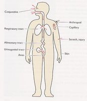
Pathogens must enter the host through specific sites known as portals of entry. Most pathogens have a preferred portal, which influences their ability to cause disease.
Mucous membranes: Includes the respiratory tract, gastrointestinal tract (enteral route), genitourinary tract, placenta, and conjunctiva of the eye.
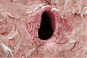
Parenteral route: Entry through broken skin, wounds, bites, injections, or punctures.
Numbers of Invading Microbes: ID50 and LD50
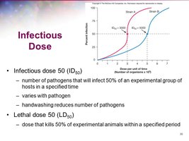
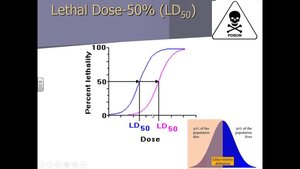
The likelihood of disease depends on the number of microbes entering the host. Two important measures are:
ID50 (Infectious Dose 50): The number of pathogens required to infect 50% of a sample population.
LD50 (Lethal Dose 50): The dose of a toxin that kills 50% of a sample population, often measured in mg/kg body weight.
Adherence
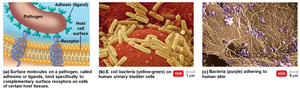
Almost all pathogens must attach to host tissues in a process called adherence (or adhesion). This is mediated by specific molecules:
Adhesins (ligands): Surface molecules on the pathogen that bind to receptors on host cells.
Structures involved: Glycocalyx, fimbriae.
Biofilms: Microbes may form biofilms, which are communities that share nutrients and enhance adherence.
How Pathogens Penetrate Host Defenses
Capsules
Some bacteria produce a glycocalyx capsule around their cell wall, which impairs phagocytosis and increases pathogenicity.
Examples: Streptococcus pneumoniae (pneumonia), Haemophilus influenzae (pneumonia and meningitis), Bacillus anthracis (anthrax), Yersinia pestis (plague).
Cell Wall Components
M protein: Resists phagocytosis (Streptococcus pyogenes).
Opa protein: Allows attachment to host cells (Neisseria gonorrhoeae).
Waxy lipid (mycolic acid): Resists digestion (Mycobacterium tuberculosis).
Enzymes
Coagulases: Coagulate fibrinogen, protecting bacteria from phagocytosis.
Kinases: Digest fibrin clots, allowing bacteria to spread.
Hyaluronidase: Digests polysaccharides that hold cells together.
Collagenase: Breaks down collagen in connective tissue.
IgA proteases: Destroy IgA antibodies, reducing immune defense.
Penetration into the Host Cell Cytoskeleton
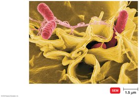
Bacteria use invasins (surface proteins) to rearrange actin filaments of the host cell cytoskeleton, causing membrane ruffling and facilitating entry.
Examples: Shigella, Listeria, Salmonella.
How Bacterial Pathogens Damage Host Cells
Using the Host's Nutrients: Siderophores
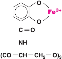
Pathogenic bacteria require iron, which is tightly bound by host proteins. Siderophores are proteins secreted by pathogens that bind iron more tightly than host molecules, facilitating iron acquisition.
Host iron-binding molecules: Transferrin, ferritin, lactoferrin, hemoglobin.
Direct Damage
Disrupts host cell function.
Uses host cell nutrients.
Produces waste products.
Multiplies in host cells, causing cell rupture.
Production of Toxins
Toxins are poisonous substances produced by microorganisms. They can cause fever, cardiovascular problems, diarrhea, and shock.
Toxigenicity: Ability to produce a toxin.
Toxemia: Presence of toxin in the host's blood.
Intoxications: Presence of toxin without microbial growth.
Exotoxins
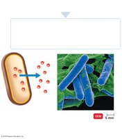
Exotoxins are proteins produced and secreted by bacteria, usually gram-positive, and are soluble in bodily fluids. They destroy host cells and inhibit metabolic functions.
Antitoxins: Antibodies against specific exotoxins.
Toxoids: Inactivated exotoxins used in vaccines.
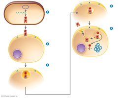
A-B Toxins
A-B toxins consist of an enzyme component (A part) and a binding component (B part). The B part binds to the host cell, and the A part exerts toxic effects.
Example: Diphtheria toxin.
Membrane-Disrupting Toxins
Leukocidins: Kill phagocytic leukocytes.
Hemolysins: Kill erythrocytes by forming protein channels.
Streptolysins: Hemolysins produced by streptococci.
Superantigens
Superantigens cause an intense immune response due to cytokine release from host cells, leading to fever, nausea, vomiting, diarrhea, shock, and death.
Genotoxins
Genotoxins damage DNA, causing mutations, disrupting cell division, and potentially leading to cancer.
Endotoxins
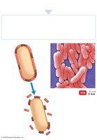
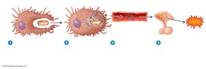
Endotoxins are the lipid A portion of lipopolysaccharides (LPS) found in the cell wall of gram-negative bacteria. They are released during bacterial multiplication and when bacteria die.
Stimulate macrophages to release cytokines.
Can cause disseminated intravascular coagulation.
Comparison of Exotoxins and Endotoxins
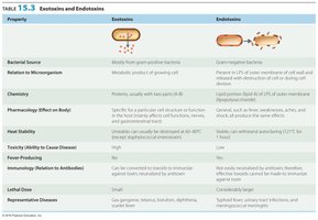
The following table summarizes the differences between exotoxins and endotoxins:
Property | Exotoxins | Endotoxins |
|---|---|---|
Bacterial Source | Mostly from gram-positive bacteria | Gram-negative bacteria |
Chemistry | Proteins, usually with two parts (A-B) | Lipid portion of LPS |
Pharmacology Effect | Specific for a particular cell structure or function | General, such as fever, weakness, aches |
Heat Stability | Unstable | Stable |
Toxicity | High | Low |
Fever Producing | No | Yes |
Immunity | Can be converted to toxoids for immunization | Not easily neutralized by antibodies |
Lethal Dose | Small | Large |
Representative Diseases | Tetanus, botulism, diphtheria | Typhoid fever, urinary tract infections |
Plasmids, Lysogeny, and Pathogenicity
Plasmids: May carry genes for toxins, antibiotic production, and enzymes.
Lysogenic conversion: Changes characteristics of a microbe due to incorporation of a prophage.
Pathogenic Properties of Viruses
Cytopathic Effects (CPE)
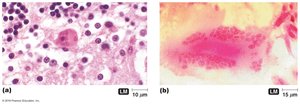
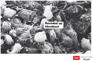
Cytopathic effects are visible changes in host cells due to viral infection. These effects include:
Stopping cell synthesis
Causing cell lysosomes to release enzymes
Creating inclusion bodies in the cytoplasm
Fusing cells to create a syncytium
Changing host cell function or inducing chromosomal changes
Inducing antigenic changes on the cell surface
Loss of contact inhibition, leading to cancer
Producing interferons to protect uninfected cells
Pathogenic Properties of Fungi, Protozoa, Helminths, and Algae
Fungi
Toxic metabolic products provoke allergic responses.
Trichothecene toxins inhibit protein synthesis.
Proteases modify host cell membranes.
Capsules prevent phagocytosis.
Ergot alkaloid toxins cause hallucinations.
Aflatoxin is a carcinogenic toxin produced by Aspergillus.
Mycotoxins (e.g., phalloidin, amanitin) are neurotoxic.
Protozoa
Presence and waste products cause symptoms.
Evade host defenses by digesting cells and tissue fluids, growing in phagocytes, and antigenic variation.
Helminths
Use host tissue for growth.
Produce large masses and waste products that cause symptoms.
Algae
Some produce neurotoxins, such as saxitoxin, causing paralytic shellfish poisoning.
Portals of Exit
Pathogens leave the host through specific portals of exit, which are generally the same as the portals of entry.
Respiratory tract: Coughing and sneezing
Gastrointestinal tract: Feces and saliva
Genitourinary tract: Urine; secretions from the penis and vagina
Skin
Blood: Arthropods that bite; needles or syringes
Key Concepts
Portals of entry and exit are critical for infection.
Pathogens adhere, penetrate or evade host defenses, and damage host tissues.
Multiple factors contribute to pathogenicity, including toxins, enzymes, and genetic elements.
