Back
BackMicrobial Mechanisms of Pathogenicity: Study Notes
Study Guide - Smart Notes
 Tailored notes based on your materials, expanded with key definitions, examples, and context.
Microbial Mechanisms of Pathogenicity
Introduction
This chapter explores how microorganisms cause disease in hosts, focusing on the mechanisms of pathogenicity, the strategies microbes use to evade host defenses, and the ways they damage host cells. Understanding these processes is essential for microbiology students to grasp the complexity of infectious diseases.
The Spread of Infection
Stages of Infection
Encounter: The initial contact between the microbe and the host.
Entry: Microorganisms enter the host through specific portals.
Spread: The pathogen disseminates within the host.
Multiplication: The pathogen increases in number.
Evasion of Host Defenses: Microbes avoid immune responses.
Damage: Pathogen causes harm to host tissues.
Outcome: Transmission to a new host or recovery/death of the host.
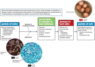
How Microorganisms Enter a Host
Key Definitions
Pathogenicity: The ability of a microorganism to cause disease.
Virulence: The degree of pathogenicity.
Pathogenesis: The process by which a microorganism causes disease.
Portals of Entry
Mucous membranes: Respiratory tract (most common), digestive canal, genital system, urinary system, conjunctiva.
Skin: Usually impenetrable unless there are cuts, abrasions, or entry via hair follicles/sweat glands.
Parenteral route: Direct deposition into tissues beneath the skin or mucous membranes (e.g., injections, bites, wounds, surgery).
Most pathogens have a preferred portal of entry; entry by other means may not cause disease.
Numbers of Invading Microbes
Infectious and Lethal Doses
ID50: Infectious dose for 50% of a sample population (measures virulence).
LD50: Lethal dose for 50% of a sample population (measures toxin potency).
Portal of Entry | ID50 for Bacillus anthracis |
|---|---|
Skin | 10–50 endospores |
Inhalation | 10,000–20,000 endospores |
Ingestion | 250,000–1,000,000 endospores |
Toxin | LD50 |
|---|---|
Botulinum | 0.03 ng/kg |
Shiga toxin | 250 ng/kg |
Staphylococcal enterotoxin | 1350 ng/kg |
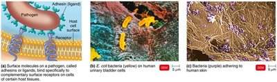
Adherence
Mechanisms of Adherence
Pathogens attach to host tissues using adhesins (ligands) that bind to specific receptors on host cells.
Examples:
Glycocalyx of Streptococcus mutans enables adherence to teeth.
Fimbriae of Actinomyces adhere to the glycocalyx of S. mutans.
Viral spikes (e.g., SARS-CoV-2) adhere to ACE2 receptors on host cells.
How Pathogens Penetrate Host Defenses
Capsules
Composed of glycocalyx around the cell wall.
Impair phagocytosis (e.g., Streptococcus pneumoniae, Haemophilus influenzae, Bacillus anthracis, Yersinia pestis).
Cell Wall Components
M protein: Resists phagocytosis (Streptococcus pyogenes).
Opa protein: Allows attachment to host cells (Neisseria gonorrhoeae).
Waxy lipid (mycolic acid): Resists digestion by phagocytes (Mycobacterium tuberculosis).
Enzymes
Coagulases: Coagulate fibrinogen, forming fibrin to protect bacteria.
Kinases: Digest fibrin clots.
Hyaluronidase: Digests hyaluronic acid, aiding spread.
Collagenase: Breaks down collagen (important in gas gangrene).
IgA proteases: Destroy IgA antibodies.
Antigenic Variation
Pathogens alter their surface antigens, rendering antibodies ineffective.
Examples: Influenza virus, Neisseria gonorrhoeae, Trypanosoma brucei gambiense.
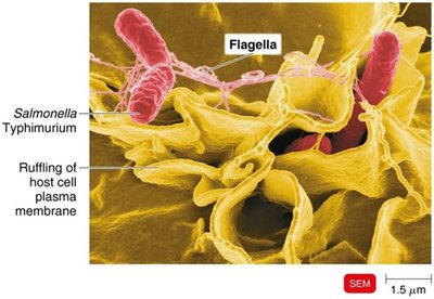
Penetration into the Host
Invasins: Surface proteins that rearrange actin filaments, causing membrane ruffling and engulfment (e.g., Shigella, Listeria).
Some bacteria use actin to move between cells.
Some survive inside phagocytes by escaping the phagosome, preventing lysosomal fusion, or tolerating low pH.
Biofilms
Biofilms resist antibiotics and disinfectants.
Involved in 65% of all infections.
Biofilm bacteria are more resistant to phagocytosis due to the extracellular polymeric substance (EPS).
How Bacterial Pathogens Damage Host Cells
Using the Host’s Nutrients: Siderophores
Pathogens secrete siderophores to bind iron more tightly than host proteins.
Can also take up host iron transport cells or kill host cells for iron.
Direct Damage
Disrupts host cell function, uses nutrients, produces waste, and causes cell rupture.
Production of Toxins
Toxins: Poisonous substances produced by microorganisms.
Cause fever, cardiovascular problems, diarrhea, and shock.
Toxigenicity: Ability to produce a toxin.
Toxemia: Presence of toxin in the blood.
Intoxications: Disease caused by toxin without microbial growth.
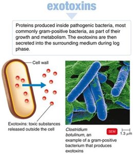
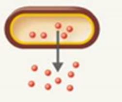
Exotoxins
Proteins produced and secreted by bacteria (mainly gram-positive, but also some gram-negative).
Soluble in body fluids, highly specific, and often highly lethal.
Antitoxins (antibodies) provide immunity; toxoids (inactivated toxins) are used in vaccines.
Enzymatic activity allows small amounts to be very harmful.
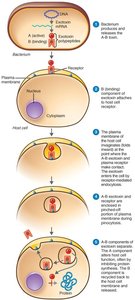
A-B Toxins and Other Exotoxins
A-B toxins: Consist of an enzyme (A) and a binding (B) component (e.g., diphtheria toxin).
Genotoxins: Damage DNA, causing mutations and possibly cancer.
Membrane-disrupting toxins: Lyse host cells by disrupting membranes (e.g., leukocidins, hemolysins, streptolysins).
Superantigens: Cause intense immune response by stimulating cytokine release, leading to fever, shock, and death.
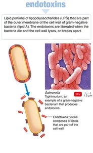
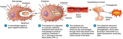
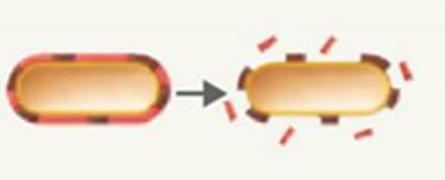
Endotoxins
Lipid A portion of lipopolysaccharides (LPS) in gram-negative bacteria.
Released during bacterial multiplication or cell death.
Stimulate macrophages to release cytokines, causing fever, chills, weakness, and shock.
Can cause disseminated intravascular coagulation (DIC) and endotoxic shock.
Detection of Endotoxins
Limulus amebocyte lysate (LAL) assay: Uses horseshoe crab blood to detect endotoxins by clot formation.
Comparison of Exotoxins and Endotoxins
Property | Exotoxins | Endotoxins |
|---|---|---|
Bacterial Source | Gram-positive and gram-negative | Gram-negative |
Relation to Microorganism | Metabolic product of growing cell | Part of LPS, released on cell death/division |
Chemistry | Proteins (A-B type) | Lipid A of LPS |
Effect on Body | Specific (cell functions, nerves, GI tract) | General (fever, aches, shock) |
Heat Stability | Unstable (destroyed at 60–80°C) | Stable (withstands 121°C) |
Toxicity | High | Low |
Fever-Producing | No | Yes |
Immunology | Can be neutralized by antitoxin, toxoids for vaccines | Not easily neutralized, no effective toxoids |
Lethal Dose | Small | Larger |
Representative Diseases | Gas gangrene, tetanus, botulism, diphtheria, scarlet fever | Typhoid fever, UTIs, meningococcal meningitis |
Plasmids, Lysogeny, and Pathogenicity
Plasmids: May carry genes for toxins, antibiotic resistance (R plasmids), and virulence factors.
Lysogenic conversion: Incorporation of a prophage can change microbial characteristics (e.g., diphtheria toxin genes).
Pathogenic Properties of Viruses
Mechanisms of Viral Pathogenicity
Intracellular location shields viruses from immune detection.
Attachment via host cell surface molecules.
Direct attack on immune system components.
Mimicry of host RNA via methylation.
Antigenic variation.
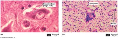
Cytopathic Effects (CPE)
Visible effects of viral infection on host cells.
Cytocidal effects: Kill host cells.
Noncytocidal effects: Cause cell damage without death.
Examples: Disruption of cell junctions, cytokine storms, inclusion bodies (e.g., Negri bodies in rabies), syncytia formation, chromosomal damage, loss of contact inhibition (cancer).
Virus (Genus) | Cytopathic Effect |
|---|---|
Poliovirus (Enterovirus C) | Cytocidal (cell death) |
Genital warts virus (Alphapapillomavirus) | Acidophilic inclusion bodies in nucleus, transformation |
Adenovirus (Mastadenovirus) | Basophilic inclusion bodies in nucleus |
Rabies (Lyssavirus) | Acidophilic inclusion bodies in cytoplasm |
CMV (Cytomegalovirus) | Acidophilic inclusion bodies in nucleus and cytoplasm |
Measles virus (Morbillivirus) | Cell fusion |
HIV (Lentivirus) | Destruction of T cells |
SARS-CoV-2 (Betacoronavirus) | Syncytia, cilia shrinkage, altered junctions |
Interferons
Alpha and beta interferons are produced by virally infected cells.
Protect neighboring cells by inhibiting viral protein synthesis and inducing apoptosis.
Some viruses can evade interferon effects.
Pathogenic Properties of Fungi, Protozoa, Helminths, and Algae
Fungi
Produce toxic metabolic products and provoke allergic responses.
Trichothecene toxins inhibit protein synthesis.
Proteases modify host cell membranes (e.g., Candida albicans).
Capsules prevent phagocytosis (Cryptococcus neoformans).
Ergot alkaloids cause hallucinations; aflatoxin (from Aspergillus) is carcinogenic; mycotoxins (e.g., phalloidin, amanitin) are neurotoxic.
Protozoa
Cause symptoms via their presence and waste products.
Evade defenses by digesting cells, growing in phagocytes, and antigenic variation (e.g., Giardia intestinalis, Toxoplasma gondii, Trypanosoma).
Helminths
Use host tissue for growth, produce large masses, and cause cellular damage.
Waste products may cause symptoms.
Algae
Some produce neurotoxins (e.g., saxitoxin) causing paralytic shellfish poisoning.
Associated with red tides (dinoflagellate Alexandrium); toxins accumulate in mollusks and affect humans who consume them.
Portals of Exit
Generally the same as portals of entry.
Respiratory tract: Coughing, sneezing.
Gastrointestinal tract: Feces, saliva.
Genitourinary tract: Urine, secretions.
Skin
Blood: Arthropod bites, needles, syringes.