 Back
BackMicrobial Mechanisms of Pathogenicity: Study Notes
Study Guide - Smart Notes
 Tailored notes based on your materials, expanded with key definitions, examples, and context.
Microbial Mechanisms of Pathogenicity
Introduction
Pathogenicity refers to the ability of a microorganism to cause disease, while virulence describes the degree of pathogenicity. Understanding how microbes invade, evade host defenses, and damage host tissues is fundamental to microbiology and infectious disease.
How Microorganisms Enter a Host
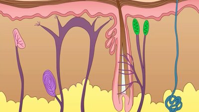
Portals of Entry
Microorganisms gain access to the body through specific portals of entry. Most pathogens have a preferred portal of entry, which is essential for their ability to cause disease.
Mucous membranes: Includes the respiratory, gastrointestinal, and genitourinary tracts, as well as the conjunctiva.
Skin: Although generally an effective barrier, some pathogens can enter through cuts, hair follicles, or sweat gland ducts.
Parenteral route: Pathogens are deposited directly into tissues when barriers are penetrated, such as by punctures, bites, or injections.
Numbers of Invading Microbes
The likelihood of disease depends on the number of invading microbes. Two important measures are:
ID50 (Infectious Dose 50): The number of microbes required to cause infection in 50% of a sample population. It measures the virulence of a microbe.
LD50 (Lethal Dose 50): The amount of toxin required to kill 50% of a sample population. It measures the potency of a toxin.
Example: Bacillus anthracis has different ID50 values depending on the portal of entry (e.g., skin: 10–50 endospores; inhalation: 10,000–20,000 endospores; ingestion: 250,000–1,000,000 endospores).
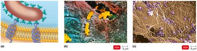
Adherence to Host Tissues
Mechanisms of Adherence
Adherence is a critical step in pathogenesis. Pathogens use surface molecules called adhesins (or ligands) to bind specifically to complementary receptors on host cells. Common adhesins include glycocalyx and fimbriae. Many microbes also form biofilms, which are communities that share nutrients and provide protection from host defenses.
Penetration or Evasion of Host Defenses
Capsules and Cell Wall Components
Some bacteria evade host defenses by producing capsules (glycocalyx layers) that impair phagocytosis. Cell wall components such as M protein, Opa protein, and mycolic acid also contribute to resistance against host immune responses.
Capsules: Found in Streptococcus pneumoniae, Haemophilus influenzae, Bacillus anthracis, and Yersinia pestis.
M protein: Resists phagocytosis (Streptococcus pyogenes).
Opa protein: Allows attachment to host cells (Neisseria gonorrhoeae).
Mycolic acid: Waxy lipid that resists digestion (Mycobacterium tuberculosis).
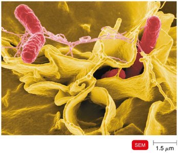
Biofilms
Biofilms protect bacteria from phagocytosis by shielding them with an extracellular polymeric substance (EPS).
Enzymes and Antigenic Variation
Bacteria produce enzymes that facilitate invasion and evasion:
Coagulases: Coagulate fibrinogen.
Kinases: Digest fibrin clots.
Hyaluronidase: Digests polysaccharides holding cells together.
Collagenase: Breaks down collagen.
IgA proteases: Destroy IgA antibodies.
Antigenic variation allows pathogens to alter their surface antigens, rendering antibodies ineffective.
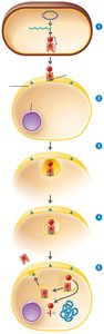
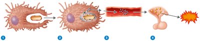
Penetration into Host Cells
Bacteria use invasins to rearrange actin filaments of the host cytoskeleton, causing membrane ruffling and facilitating entry. Some bacteria, such as Shigella and Listeria, use actin to move between cells. Survival inside phagocytes is achieved by escaping the phagosome, preventing lysosomal fusion, or surviving in low pH environments.
How Bacterial Pathogens Damage Host Cells
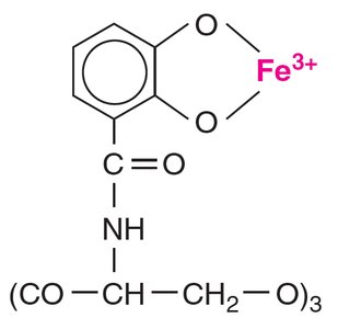
Using the Host’s Nutrients: Siderophores
Iron is essential for bacterial growth. Pathogens secrete siderophores, proteins that bind iron more tightly than host cells, allowing bacteria to acquire this vital nutrient.
Direct Damage
Bacteria can directly damage host cells by disrupting cell function, using host nutrients, producing waste products, and causing cell rupture through multiplication.
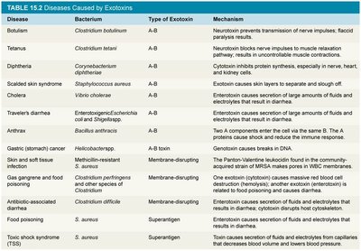
Production of Toxins
Toxins are poisonous substances produced by microorganisms that can cause fever, cardiovascular problems, diarrhea, and shock. Key terms include:
Toxigenicity: Ability to produce toxins.
Toxemia: Presence of toxin in the blood.
Intoxications: Disease caused by toxins without microbial growth.
Exotoxins
Exotoxins are proteins secreted by bacteria (mainly gram-positive) that destroy host cells or inhibit metabolic functions. They are highly specific and potent. Antitoxins are antibodies against exotoxins, and toxoids are inactivated exotoxins used in vaccines.
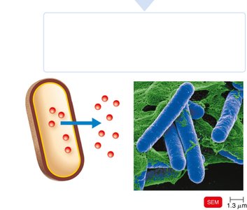
Types of Exotoxins
A-B toxins: Consist of an active (A) component and a binding (B) component (e.g., diphtheria toxin).
Genotoxins: Damage DNA, causing mutations and potentially cancer.
Membrane-disrupting toxins: Lyse host cells by disrupting plasma membranes (e.g., leukocidins, hemolysins, streptolysins).
Superantigens: Cause intense immune responses by stimulating T cells to release cytokines, leading to fever, shock, and death.
Endotoxins
Endotoxins are the lipid A portion of lipopolysaccharides (LPS) found in the outer membrane of gram-negative bacteria. They are released during bacterial multiplication and cell lysis, causing fever and potentially disseminated intravascular coagulation.
Detection of Endotoxins
The Limulus amebocyte lysate (LAL) assay uses horseshoe crab blood to detect endotoxins, as amebocytes lyse in their presence, forming a clot.
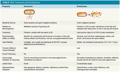
Comparison of Exotoxins and Endotoxins
Property | Exotoxins | Endotoxins |
|---|---|---|
Bacterial Source | Gram-positive and gram-negative bacteria | Gram-negative bacteria |
Chemistry | Proteins, usually with two parts (A-B) | Lipid A (LPS) of outer membrane |
Heat Stability | Unstable (destroyed at 60–80°C) | Stable (withstands autoclaving) |
Toxicity | High | Low |
Fever-Producing | No | Yes |
Representative Diseases | Gas gangrene, tetanus, botulism, diphtheria, scarlet fever | Typhoid fever, urinary tract infections, meningococcal meningitis |
Plasmids, Lysogeny, and Pathogenicity
Plasmids may carry genes for toxins, antibiotic resistance, and enzymes. Lysogenic conversion occurs when a bacteriophage integrates into the bacterial genome, potentially turning harmless bacteria into pathogens (e.g., E. coli).
Pathogenic Properties of Viruses
Cytopathic Effects (CPE)
Viruses cause visible effects on infected cells, known as cytopathic effects. These include:
Stopping cell synthesis
Causing lysosomal enzyme release
Creating inclusion bodies
Fusing cells to form syncytia
Inducing chromosomal changes
Loss of contact inhibition (leading to cancer)
Interferons
Alpha and beta interferons are produced by virally-infected cells and protect neighboring cells by inhibiting viral protein synthesis and inducing apoptosis in infected cells.
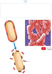
Portals of Exit
Mechanisms of Exit
Pathogens leave the host through specific portals of exit, which are generally the same as the portals of entry. These include:
Respiratory tract: Coughing and sneezing
Gastrointestinal tract: Feces and saliva
Genitourinary tract: Urine and genital secretions
Skin
Blood: Via arthropod bites or contaminated needles
Summary Table: Key Mechanisms of Pathogenicity
Portals of entry
Adherence to host tissues
Penetration or evasion of host defenses
Damage to host cells (direct damage, toxins, siderophores)
Portals of exit
Key Concept: The balance between host defenses and microbial pathogenicity determines the outcome of infection. Understanding these mechanisms is essential for disease prevention and treatment.
