 Back
BackMicrobial Metabolism: Foundations and Pathways
Study Guide - Smart Notes
 Tailored notes based on your materials, expanded with key definitions, examples, and context.
Microbial Metabolism
Defining the Requirements for Life
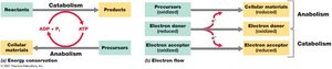
Microbial metabolism encompasses all biochemical reactions necessary for life, including both energy-releasing and energy-synthesizing processes. These reactions are categorized as catabolism (breaking down molecules to release energy) and anabolism (building cellular material using energy). Metabolic processes rely on the transfer of electrons from electron donors to electron acceptors, and cells conserve energy by converting it into forms that can perform cellular work, primarily through the generation of adenosine triphosphate (ATP).
Catabolism: Energy-releasing metabolic reactions.
Anabolism: Energy-consuming metabolic reactions that synthesize cellular components.
ATP: The universal energy currency in cells, produced during catabolic reactions and consumed during anabolic reactions.
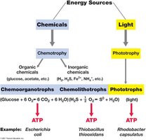
Energy Classes of Microorganisms
Microorganisms are classified based on their energy and carbon sources. These classifications help in understanding their ecological roles and metabolic diversity.
Chemoorganotrophs: Conserve energy from organic chemicals.
Chemolithotrophs: Oxidize inorganic compounds (e.g., H2, H2S, NH4+).
Phototrophs: Convert light energy into ATP.
Heterotrophs: Obtain carbon from organic compounds.
Autotrophs: Obtain carbon from CO2.
Catalysis and Enzymes
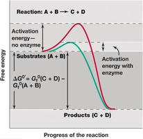
Activation Energy and Catalysis
For a chemical reaction to occur, reactant molecules must overcome an energy barrier known as activation energy. Catalysts, including biological catalysts called enzymes, lower this barrier, increasing the rate of reaction without being consumed or altering the reaction's equilibrium.
Activation Energy: Minimum energy required for molecules to become reactive.
Catalyst: Substance that facilitates a reaction, lowers activation energy, and increases reaction rate.
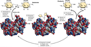
Enzyme Structure and Function
Enzymes are highly specific biological catalysts, typically proteins encoded by DNA (some are RNA). The active site of an enzyme binds the substrate, and the enzyme's shape determines its specificity. Extreme temperatures or pH can denature enzymes, affecting their function.
Active Site: Region of the enzyme where substrate binding and catalysis occur.
Substrate Specificity: Enzymes are highly specific to their substrates due to the shape of the active site.
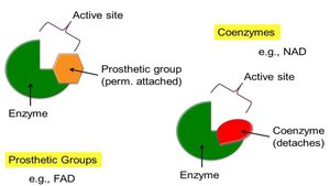
Enzyme Cofactors: Prosthetic Groups and Coenzymes
Many enzymes require non-protein molecules for catalysis. Prosthetic groups are tightly and permanently bound, while coenzymes are loosely bound and often derived from vitamins.
Prosthetic Groups: Covalently and permanently attached (e.g., heme in cytochromes).
Coenzymes: Loosely attached, often vitamin derivatives (e.g., NAD).
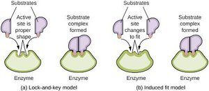
Models of Enzyme-Substrate Interaction
Enzyme catalysis depends on substrate binding and positioning relative to catalytically active amino acids. Two main models describe this interaction: the lock-and-key model and the induced fit model.
Lock-and-Key Model: Active site is a perfect fit for the substrate.
Induced Fit Model: Active site changes shape to fit the substrate upon binding.
Energy-Rich Compounds and Storage
Long-Term Energy Storage
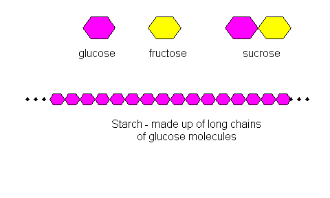
Microorganisms store energy in the form of insoluble polymers that can be oxidized to generate ATP. These storage compounds differ between prokaryotes and eukaryotes.
Prokaryotes: Glycogen (polyglucose), poly-β-hydroxybutyrate, elemental sulfur.
Eukaryotes: Starch (polyglucose), lipids (simple fats).
Glycolysis and Fermentation
Pathways of Energy Conservation
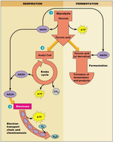
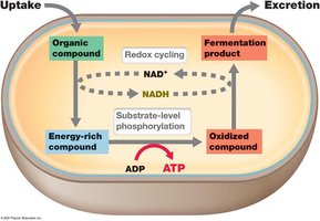
Chemoorganotrophs conserve energy through two main pathways: fermentation and respiration. Fermentation is anaerobic, while respiration can be aerobic or anaerobic, depending on the electron acceptor.
Fermentation: Anaerobic catabolism; organic compounds donate and accept electrons.
Respiration: Aerobic or anaerobic catabolism; donor is oxidized with O2 or another compound as electron acceptor.
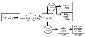
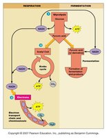
Glycolysis (Embden–Meyerhof–Parnas Pathway)
Glycolysis is a common pathway for glucose catabolism, producing two ATP molecules via substrate-level phosphorylation. Glucose can be fermented (without O2) or respired (with O2).
Substrate-Level Phosphorylation: Transfer of a phosphate group from an organic compound to ADP, forming ATP.
Fermentation Products and Organisms
Fermentation leads to various end products depending on the organism and substrate. Common products include ethanol, lactic acid, and butyric acid.
Type | Product(s) | Organisms |
|---|---|---|
Alcoholic | 2 ethanol + 2 CO2 | Yeast, Zymomonas |
Homolactic | 2 lactate | Streptococcus, some Lactobacillus |
Heterolactic | lactate + ethanol + CO2 | Leuconostoc, Oenococcus |
Mixed acid | lactate + acetate + succinate + ethanol + CO2 + H2 | Enteric bacteria |
Butyric acid | butyrate + acetate + CO2 + H2 | Clostridium |
Butanol/Acetone | butanol + acetone + CO2 + H2 | Clostridium acetobutylicum |
Caproate/Butyrate | caproate + butyrate + CO2 + H2 | Clostridium kluyveri |
Acetogenic | acetate + CO2 + H2 | Clostridium aceticum |
Electron Transport Chain (ETC) and Energy Conservation
Electron Donors and Acceptors
In eukaryotes, NADH is the primary electron donor in the ETC, with molecular oxygen as the final electron acceptor. In prokaryotes, a variety of electron donors and acceptors are used, allowing for metabolic diversity.
NADH: Key electron donor in eukaryotic ETC.
O2: Final electron acceptor in aerobic respiration.
Prokaryotes: Can use nitrate, sulfate, CO2, and other compounds as electron acceptors.
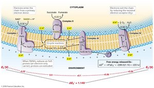
Electron Transport and the Proton Motive Force
The electron transport system is located in the cytoplasmic membrane of prokaryotes and the mitochondrial membrane of eukaryotes. As electrons are transferred, protons are released outside the membrane, generating a proton motive force (PMF)—a combination of a pH gradient and an electrochemical potential. This PMF drives ATP synthesis via ATPase.
Proton Motive Force: Electrochemical gradient generated by proton movement across the membrane.
ATP Synthesis: Most ATP is produced during electron transport.
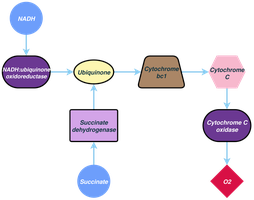
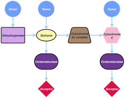
Electron Carriers in the ETC
Electron transport systems involve various carriers, including NADH dehydrogenases, flavoproteins, iron–sulfur proteins, cytochromes, and quinones. Cytochromes contain heme groups, while quinones are nonprotein carriers that transfer electrons within the membrane.
Cytochromes: Proteins with heme groups that accept and donate electrons.
Quinones: Nonprotein molecules that shuttle electrons and protons.

Options for Energy Conservation
Anaerobic Respiration
Some microorganisms use electron acceptors other than oxygen, such as nitrate, ferric iron, sulfate, carbon dioxide, or organic compounds. Anaerobic respiration conserves less energy than aerobic respiration but still generates a proton motive force and uses ATPase.
Electron Acceptors: Nitrate (NO3–), ferric iron (Fe3+), sulfate (SO42–), CO2, fumarate.
Energy Conservation: Less efficient than aerobic respiration.
Chemolithotrophy
Chemolithotrophs use inorganic chemicals as electron donors, such as hydrogen sulfide, hydrogen gas, ferrous iron, and ammonium. These organisms are typically autotrophic, using CO2 as a carbon source, and their metabolism is often aerobic.
Electron Donors: H2S, H2, Fe2+, NH4+.
Autotrophy: Use CO2 for biosynthesis.
Phototrophy
Phototrophs use light as an energy source, synthesizing ATP via photophosphorylation. Photoautotrophs use ATP and CO2 for biosynthesis, while photoheterotrophs use ATP and organic carbon.
Photoautotrophs: ATP + CO2 for biosynthesis.
Photoheterotrophs: ATP + organic carbon for biosynthesis.
Biosynthesis of Cellular Components
Sugars and Polysaccharides
Prokaryotic polysaccharides are synthesized from glucose derivatives, such as uridine diphosphoglucose (UDPG). Gluconeogenesis allows synthesis of glucose from non-carbohydrate precursors, and pentose sugars are produced via the pentose pathway for nucleic acid synthesis.
UDPG: Precursor for important polysaccharides.
Gluconeogenesis: Synthesis of glucose from phosphoenolpyruvate.
Pentose Pathway: Produces 5-carbon sugars for DNA/RNA.
Amino Acids: Protein Monomers
Amino acid biosynthesis involves multistep pathways, with carbon skeletons derived from glycolysis or the citric acid cycle. The amine group is incorporated from ammonia and transferred by specific enzymes.
Carbon Skeletons: Derived from glycolysis or citric acid cycle intermediates.
Amine Group: Incorporated by glutamine dehydrogenase or glutamine synthetase.
Fatty Acids and Lipids
Fatty acid biosynthesis varies with species and temperature. Lower temperatures favor shorter, unsaturated fatty acids, while higher temperatures favor longer, saturated fatty acids. In Bacteria and Eukarya, lipids are formed by adding fatty acids to glycerol, while Archaea use phytanyl side chains. All membranes have a hydrophobic interior and hydrophilic surfaces.
Bacteria/Eukarya: Fatty acids added to glycerol.
Archaea: Lipids contain phytanyl side chains.
Membrane Structure: Polar groups create hydrophobic and hydrophilic regions.
