 Back
BackMicrobial Pathogenesis: Host-Pathogen Interactions and Virulence Mechanisms
Study Guide - Smart Notes
 Tailored notes based on your materials, expanded with key definitions, examples, and context.
Microbial Pathogenesis
Host–Pathogen Interactions
The outcome of an infection is determined by the dynamic interplay between the pathogen's survival strategies and the host's immune responses. Both parties continuously adapt, resulting in a complex relationship that shapes disease progression.
Pathogen Strategies: Pathogens evolve mechanisms to invade, survive, and replicate within the host.
Host Responses: The host deploys immune defenses to detect and eliminate pathogens.
Adaptation: Both host and pathogen undergo genetic and phenotypic changes to outcompete each other.
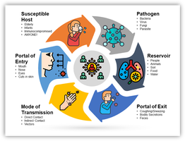
Infection Cycle
Pathogens follow structured cycles to maintain their populations, often requiring specific host environments to complete their life cycles.
Establishment: Breaching physical barriers, moving to infection sites, and becoming established are essential steps.
Transmission: Pathogen success is measured by its ability to spread to new hosts without causing premature host death.
Host Specificity: Pathogens may be specialists (infecting one host species) or generalists (infecting multiple species).
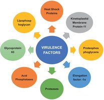
Virulence Factors
Definition and Core Functions
Virulence factors are molecular tools that enable microbes to cause damage and survive within the host environment. They facilitate host cell invasion, nutrient acquisition, and subversion of host signaling pathways.
Host Cell Invasion: Enables pathogens to penetrate and colonize host tissues.
Nutrient Acquisition: Mechanisms such as iron scavenging are critical for microbial survival.
Subversion of Host Signaling: Pathogens manipulate host cell processes to evade immune responses.
Genetic Basis and Environmental Sensing
Pathogenicity Islands: Virulence genes are often located on discrete DNA segments that can be transferred between microbes.
Environmental Sensing: Pathogens switch specific virulence genes on or off in response to host environmental cues.
Immunopathogenesis
Host Immune Response and Disease Severity
Immunopathogenesis refers to how the host's immune response contributes to disease symptoms and tissue damage.
Bystander Damage: Inflammation intended to clear pathogens can destroy healthy tissues.
Autoimmunity: Molecular mimicry may cause the immune system to attack self-tissues.
Outcome Determinant: Disease severity is often a balance between pathogen damage and host hypersensitivity.
Pathogen Evolution
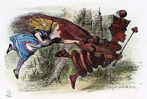
Evolutionary Arms Race
Pathogens and hosts are engaged in an "evolutionary arms race," known as the Red Queen hypothesis, where each adapts to the other's advances.
Antigenic Escape: Pathogens evolve mutations to avoid recognition by host T cells and antibodies.
Virulence Trade-offs: Evolution favors the level of virulence that maximizes transmission, not necessarily harmlessness.
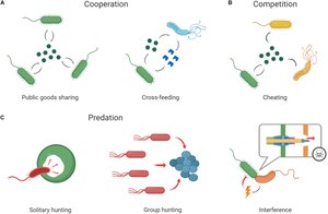
Microbiota Competition
The presence of resident microbiota forces pathogens to evolve rapidly to compete for resources and niche space.
Microbial Attachment: First Contact
Adhesion and Biofilm Formation
Pathogens rely on adhesion and biofilm formation to stay within the host and protect themselves from external threats.
Adhesion: Specialized surface molecules called adhesins bind to specific host cell receptors.
Biofilm Formation: Microbes transition from a planktonic state to a sessile, communal lifestyle within an extracellular matrix.
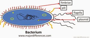
Pathogen Adhesion to Host Cells
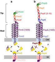
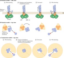
Pili and Fimbriae: Hair-like appendages with adhesin proteins at their tips match host receptors.
Type 1 Pili: Found in E. coli, bind to mannose receptors in the urinary tract.
Type IV Pili: Enable "twitching motility" for optimal colonization.
Signal Transduction
Adhesion triggers signaling in both pathogen and host, activating virulence genes and inducing host cell internalization.

The Role of Biofilms in Infection
Biofilm Life Cycle
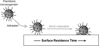
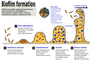
Biofilms are structured communities of microorganisms enclosed in a self-produced extracellular polymeric substance (EPS) matrix. They transition from planktonic to sessile states, forming complex structures.
Reversible Attachment: Weak adhesion via physical forces and appendages.
Irreversible Attachment: EPS matrix production anchors the microcolony.
Maturation: Growth into three-dimensional structures with water channels.
Dispersion: Environmental cues trigger detachment and colonization of new sites.


Clinical Significance & Persistence
Antibiotic Tolerance: Biofilm bacteria are 100–1,000 times more resistant to antibiotics.
Immune Evasion: The matrix shields bacteria from phagocytosis and neutralizes antibodies.
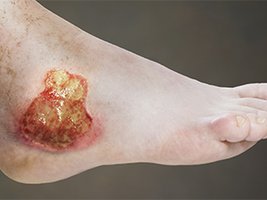
Chronic Inflammation: Persistent biofilms lead to chronic immune activation and tissue damage.
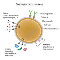
Toxins Subvert Host Functions
Microbial Toxins
Microbial toxins are specialized proteins or molecules that disrupt host cell functions, creating a favorable environment for the pathogen.
Exoenzymes: Digest host tissues to allow pathogen spread and access to nutrients.
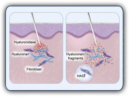
Hyaluronidase and Collagenase: Break down extracellular matrix, facilitating invasion.
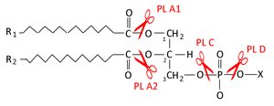
Phospholipases: Degrade host cell membranes, causing cell lysis and nutrient release.
Disarming the Immune System
Leukocidins: Target and kill white blood cells.
Superantigens: Trigger a non-specific cytokine storm, causing systemic chaos.
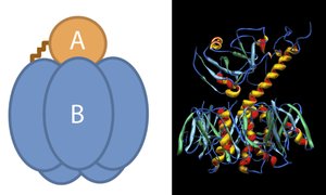
Hijacking Intracellular Signaling (A-B Toxins)
Many potent toxins use a two-part (A-B) structure to take over host cells from the inside.
B Component: Binds to specific surface receptors, facilitating entry.
A Component: Modifies host proteins to alter their function.

Examples of A-B Toxins
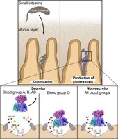
Cholera: A-subunit forces intestinal cells to pump out electrolytes and water, causing diarrhea and facilitating transmission.
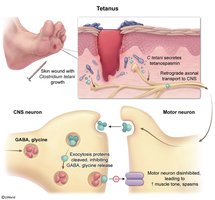
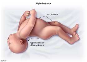
Tetanus/Botulism: Toxins block neurotransmitter release, causing paralysis.
Direct Cytotoxicity and Pore Formation
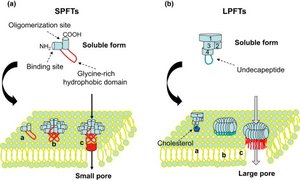
Pore-Forming Toxins (PFTs): Proteins assemble into rings on host membranes, creating holes and causing cell death by osmotic shock.
The Evolution of Toxins
Toxins are evolved to optimize pathogen survival and transmission. For example, toxins that induce coughing or sneezing facilitate pathogen spread to new hosts.
Summary Table: Virulence Factors and Their Functions
Virulence Factor | Function | Example |
|---|---|---|
Adhesins | Attachment to host cells | Pili in E. coli |
Biofilm Matrix | Protection from immune system and antibiotics | EPS in Pseudomonas aeruginosa |
Exoenzymes | Breakdown of host tissues | Hyaluronidase, Collagenase |
A-B Toxins | Hijacking host cell signaling | Cholera, Diphtheria, Tetanus toxins |
Pore-Forming Toxins | Direct cytotoxicity | Streptolysin O |
Superantigens | Immune system disruption | TSST-1 in Staphylococcus aureus |
Additional info: Academic context was added to clarify mechanisms, provide definitions, and expand on examples for completeness.
