 Back
BackMicrobial Pathogenesis, Human Microbiome, and Virulence Factors
Study Guide - Smart Notes
 Tailored notes based on your materials, expanded with key definitions, examples, and context.
Microbial Symbiosis with Humans
Overview of the Human Microbiome
The human body is inhabited by a vast array of microorganisms, collectively known as the microbiota. These include bacteria, archaea, fungi, and viruses, which colonize various anatomical sites such as the mouth, nasal cavity, throat, stomach, intestines, urogenital tract, and skin. The combination of the host and its associated microbes is referred to as a host-microbiome supraorganism. The composition of the microbiota is influenced by factors such as diet, activities, health status, birth mode, and antibiotic use.
Microbiome: The total genetic content of all microorganisms in a particular environment.
Microbiota: The community of microorganisms themselves.
Flora: An older term for microbiota, often used interchangeably.
Key anatomical sites and predominant microbes:
Skin: Propionibacterium, Actinobacteria, Staphylococcus
Saliva: Streptococcus
Urogenital tract: Lactobacillus
GI tract: Bacteroidetes
The microbiome is established early in life, with significant influences from birth mode (vaginal vs. cesarean) and feeding (breast milk vs. formula). By age 2-3, the gut microbiome stabilizes and becomes unique to each individual.
Microbial Habitats and Diversity
Skin: Different regions (dry, moist, sebaceous) have distinct microbial communities. Actinobacteria and Firmicutes are predominant.
Respiratory Tract: The upper tract harbors more diverse microbes than the lower tract. The lower tract is relatively sterile in healthy individuals.
Dental Plaque: A highly organized biofilm with early colonizers like Corynebacterium and Streptococcus. Veillonella metabolizes lactic acid produced by Streptococcus.
Urogenital Tract: The bladder and kidneys are sterile; the urethra and cervix contain abundant bacteria. Lactobacillus maintains low pH, suppressing pathogens.
GI Tract: Features a large surface area and varying pH/O2 levels. Helicobacter pylori colonizes the stomach; the colon is anaerobic and densely populated.
Microbial Community Imbalance and Disease
Dysbiosis: An imbalance in the microbiome, often leading to disease or increased susceptibility to infection.
Clostridioides difficile (C. diff): Overgrows after antibiotic treatment disrupts normal flora, leading to infection. Probiotics and fecal microbiota transplantation (FMT) can help restore balance.
Microbial Pathogenesis and Virulence
General Steps of Bacterial Pathogenesis
To cause disease, bacteria must complete several steps:
Exposure: Entry into the host (e.g., ingestion, inhalation).
Adherence: Attachment to host tissues via structures like fimbriae, pili, or adhesins.
Invasion: Entry into host cells or tissues, often by disrupting host cell structures.
Multiplication: Growth and replication, often requiring nutrient acquisition (e.g., iron via siderophores).
Toxicity: Production of toxins (exotoxins, endotoxins) that damage host tissues.
Invasiveness: Ability to spread within host tissues (e.g., via capsules).
Exit: Leaving the host to infect new individuals (e.g., via diarrhea).
Example: Salmonella typhi
Exposure: Food-borne transmission
Adherence: Type 1 fimbriae
Invasion: Disrupts actin filaments, induces membrane ruffling
Multiplication: Siderophores for iron acquisition
Toxicity: Exotoxins, LPS (endotoxin), cytotoxins
Invasiveness: Vi capsule
Exit: Enterotoxin-induced diarrhea
Virulence Factors
Virulence factors are molecules produced by pathogens that enable them to infect, evade the immune system, and cause disease. Major types include:
Exoenzymes: Enzymes that degrade host tissues (e.g., hyaluronidase, coagulase, streptokinase).
Toxins: Substances that directly damage host cells (exotoxins, endotoxins).
Capsules: Polysaccharide layers that protect bacteria from immune detection.
Adhesins: Surface proteins that mediate attachment to host cells.
Siderophores: Molecules that scavenge iron from the host (not always considered a virulence factor).
Enzymes as Virulence Factors
Hyaluronidase: Breaks down hyaluronic acid in the extracellular matrix, allowing bacteria to invade deeper tissues.
Coagulase: Induces blood clotting, protecting bacteria from immune cells (Staphylococcus).
Streptokinase: Dissolves blood clots, facilitating bacterial spread (Streptococcus).
IgA1 Protease: Degrades IgA1 antibodies, helping bacteria evade mucosal immunity (e.g., Neisseria, Haemophilus).
Toxins
Exotoxins: Secreted proteins that cause specific damage (e.g., enterotoxins affecting the intestines).
Endotoxins: Lipopolysaccharide (LPS) components of Gram-negative bacteria; Lipid A is the toxic part. Systemic exposure can cause septic shock.
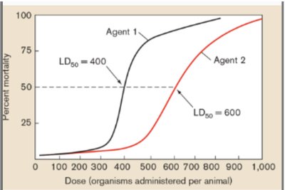
Measuring Virulence: LD50 and Attenuation
Virulence is often quantified by the lethal dose 50 (LD50), the number of organisms required to kill 50% of a host population. Lower LD50 indicates higher virulence.
Attenuation refers to the loss of virulence, often used in vaccine development. Attenuated strains can stimulate immunity without causing disease.
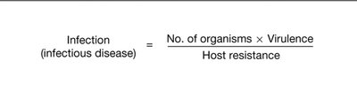
Equation for Infectious Disease
The likelihood of infection depends on the number of organisms, their virulence, and the host's resistance:
Host-Microbe Interactions and Disease Determinants
Determinants of Disease
Not all microbes are pathogenic; disease depends on both microbial properties (e.g., presence of virulence genes) and host factors (e.g., immune status).
Example: Escherichia coli is normally harmless in the gut but can cause disease if it acquires toxin genes or infects immunocompromised hosts.
Portals of Entry and Exit
Common entry points: ingestion, inhalation, mucous membranes, vertical transmission (mother to child).
Exit strategies: often involve symptoms that facilitate spread (e.g., diarrhea, coughing).
Colonization and Immune Evasion
Bacteria often colonize mucous membranes, forming biofilms and evading immune responses via capsules or toxins (e.g., leukocidins).
Siderophores are critical for iron acquisition but are not considered classic virulence factors.
Microbiome and Human Health
Metabolic and Immune Contributions
Microbiota produce vitamins (e.g., B, K), train the immune system, and modulate inflammation via molecules like LPS.
Disruption of the microbiome (e.g., by antibiotics) can lead to disease, highlighting the importance of microbial balance.
Research and Applications
Fecal microbiota transplantation (FMT) is used to treat recurrent C. difficile infections.
Microbial modulation and engineered bacteria are emerging strategies in cancer immunotherapy.
Summary Table: Key Virulence Factors and Their Functions
Virulence Factor | Produced By | Function |
|---|---|---|
Hyaluronidase | Streptococcus | Degrades extracellular matrix, aids tissue invasion |
Coagulase | Staphylococcus | Induces clotting, protects from immune cells |
Streptokinase | Streptococcus | Dissolves clots, facilitates spread |
IgA1 Protease | Neisseria, Haemophilus, Strep | Degrades IgA1, evades mucosal immunity |
Exotoxin | Various | Damages host cells/tissues |
Endotoxin (LPS) | Gram-negative bacteria | Triggers inflammation, can cause shock |
Siderophore | Various bacteria | Scavenges iron from host |
Additional info:
Not all infections lead to disease; infection refers to colonization, while disease involves tissue damage and symptoms.
Quorum sensing is a mechanism by which bacteria coordinate gene expression based on population density, often regulating virulence factor production.
Antimicrobial enzymes (e.g., lysozyme in saliva) limit microbial growth at certain body sites.
