 Back
BackMicrobial Pathogenesis: Mechanisms of Infection and Host Damage
Study Guide - Smart Notes
 Tailored notes based on your materials, expanded with key definitions, examples, and context.
Microbial Pathogenesis
Overview of Pathogenesis
Microbial pathogenesis refers to the process by which microorganisms cause disease in a host. To establish infection, pathogens must enter the host, adhere to tissues, evade or penetrate host defenses, and inflict damage.
Portals of Entry: Mucous membranes, skin, and parenteral routes are the main entry points for pathogens.
Key Steps: Entry, adherence, invasion, evasion of host defenses, and host damage.
Portals of Entry
Mucous Membranes
Mucous membranes line the respiratory, gastrointestinal (GI), and genitourinary tracts, as well as the conjunctiva. They are the most common portals of entry for pathogens.
Respiratory Tract: Easiest and most frequently used portal. Pathogens are inhaled via droplets or dust. Examples: Common cold, pneumonia, tuberculosis, influenza, measles.
Gastrointestinal Tract: Pathogens enter via contaminated food, water, or fingers. Most are destroyed by stomach acid, but some survive and cause disease. Examples: Poliomyelitis, hepatitis A, typhoid fever, amebic dysentery, giardiasis, shigellosis, cholera.
Genitourinary Tract: Portal for sexually transmitted diseases (STDs). Examples: HIV, genital warts, chlamydia, herpes, syphilis, gonorrhea.
Conjunctiva: Lines eyelids and covers the white of the eyes. Examples: Conjunctivitis, trachoma, ophthalmia neonatorum.
Skin
The skin is a major barrier to infection. Most microorganisms cannot penetrate unbroken skin, but some can enter through hair follicles, sweat gland ducts, or by boring through intact skin (e.g., hookworm larvae).
Diseases: Conjunctivitis, trachoma, ophthalmia neonatorum.
Parenteral Route
Pathogens can be deposited directly into tissues beneath the skin or mucous membranes via punctures, injections, bites, cuts, wounds, or surgery.
Examples: Infections following injuries, surgery, or insect bites.
Preferred Portal of Entry
Many pathogens must enter through a specific portal to cause disease. Entry through a non-preferred portal may not result in infection.
Numbers of Invading Microbes
The likelihood of disease increases with the number of invading pathogens. Two important measures are:
ID50: Infectious dose for 50% of a sample population.
LD50: Lethal dose for 50% of a sample population.
Adherence
Mechanisms of Adherence
Adherence is the process by which pathogens attach to host tissues, a critical step in infection. This is mediated by surface molecules called adhesins or ligands that bind to specific receptors on host cells.
Adhesins: Often glycoproteins or lipoproteins found on glycocalyx, pili, fimbriae, or flagella.
Host Receptors: Typically sugars such as mannose.
Prevention: Altering adhesins or receptors can prevent infection.
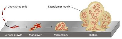
Biofilms
Microbes can form biofilms—complex communities of microorganisms adhering to surfaces and encased in an exopolymer matrix. Biofilms are resistant to disinfectants and antibiotics and are implicated in many human infections.
Examples: Dental plaque, algae on swimming pool walls, shower door scum.
Penetration of Host Defenses
Capsules
Some bacteria produce a glycocalyx that forms a capsule around the cell wall, increasing virulence by resisting phagocytosis.
Examples: Streptococcus pneumoniae, Klebsiella pneumoniae, Haemophilus influenzae, Bacillus anthracis.
Cell Wall Components
Certain cell wall components enhance virulence:
M protein: Found in Streptococcus pyogenes; resists phagocytosis and aids attachment.
Opa protein: In Neisseria gonorrhoeae; aids attachment and invasion of host cells.
Mycolic acid: Waxy lipid in Mycobacterium tuberculosis; resists digestion by phagocytes.
Enzymes (Exoenzymes)
Some bacteria secrete enzymes that aid in invasion and evasion of host defenses:
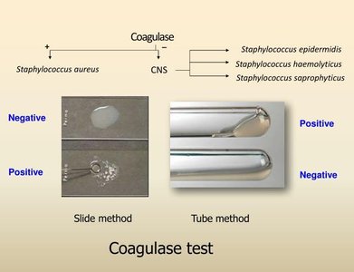
Coagulases: Clot fibrinogen in blood, protecting bacteria from phagocytosis.
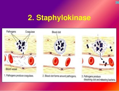
Kinases: Digest fibrin clots, allowing spread of infection.
Hyaluronidase: Hydrolyzes hyaluronic acid in connective tissue, aiding spread.
Collagenase: Breaks down collagen, facilitating tissue invasion (e.g., gas gangrene).
IgA proteases: Destroy IgA antibodies, aiding evasion of immune response.
Antigenic Variation
Some pathogens can alter their surface antigens, evading recognition by host antibodies. This allows persistent or recurrent infections.
Examples: Neisseria gonorrhoeae, influenza virus.

Penetration into Host Cell Cytoskeleton
Certain bacteria (e.g., Salmonella, E. coli) produce invasins that rearrange actin filaments, causing membrane ruffling and engulfment of the microbe by the host cell.
How Bacterial Pathogens Damage Host Cells
Mechanisms of Host Damage
Using the host's nutrients (e.g., iron via siderophores)
Causing direct damage at the site of infection
Producing toxins that affect distant sites
Inducing hypersensitivity reactions
Using Host Nutrients: Siderophores
Pathogens secrete siderophores to scavenge iron from host iron-binding proteins (e.g., lactoferrin, transferrin, ferritin, hemoglobin), which is essential for bacterial growth.
Direct Damage
Pathogens can damage host cells by using them for nutrients and producing waste products, often leading to cell rupture.
Toxins
Toxins are poisonous substances produced by certain microbes, often the primary factor in disease. They can be classified as exotoxins or endotoxins.
Toxigenicity: The ability to produce toxins.
Toxemia: Presence of toxins in the blood.
Exotoxins
Exotoxins are proteins secreted by bacteria (both Gram-positive and Gram-negative) that are highly specific and potent. They are often encoded by plasmids or phages and can be neutralized by antitoxins.
Types: A-B toxins, membrane-disrupting toxins, superantigens.
Toxoids: Inactivated exotoxins used in vaccines (e.g., diphtheria, tetanus).
Types of Exotoxins
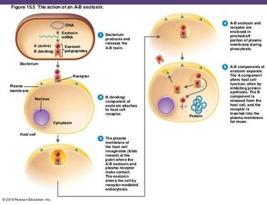
A-B Toxins: Consist of an active (A) component and a binding (B) component. Example: Diphtheria toxin.
Membrane-Disrupting Toxins: Cause cell lysis by disrupting plasma membranes.
Leukocidins: Kill phagocytic leukocytes (produced by staphylococci, streptococci).
Hemolysins: Destroy erythrocytes.
Superantigens: Bacterial proteins that provoke intense immune responses by nonspecifically activating T cells.
Specific Exotoxins
Diphtheria Toxin: Inhibits protein synthesis in eukaryotic cells (Corynebacterium diphtheriae).
Erythrogenic Toxins: Damage capillaries, causing red skin rash (scarlet fever, Streptococcus pyogenes).
Botulinum Toxin: Blocks nerve impulses, causing flaccid paralysis (Clostridium botulinum).
Tetanus Toxin: Blocks relaxation pathway, causing muscle spasms (Clostridium tetani).
Vibrio Enterotoxin: Causes secretion of fluids and electrolytes, leading to diarrhea (Vibrio cholerae).
Staphylococcal Enterotoxin: Superantigen affecting intestines (Staphylococcus aureus).
Endotoxins
Endotoxins are part of the outer membrane of Gram-negative bacteria (specifically, Lipid A of lipopolysaccharide, LPS). They are released upon bacterial cell death and lysis, causing generalized symptoms such as fever, shock, and sometimes death.
Pyrogenic Response: Endotoxins stimulate macrophages to release cytokines (e.g., IL-1, TNF-α), which induce fever by acting on the hypothalamus.
Detection: Limulus amebocyte lysate (LAL) assay is used to detect endotoxins in medical materials.
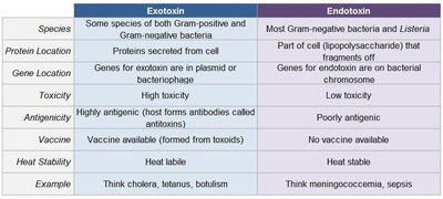
Comparison of Exotoxins and Endotoxins
Exotoxin | Endotoxin | |
|---|---|---|
Species | Some Gram-positive and Gram-negative | Most Gram-negative, some Listeria |
Protein Location | Secreted from cell | Part of cell wall (LPS) |
Gene Location | Plasmid or bacteriophage | Bacterial chromosome |
Toxicity | High | Low |
Antigenicity | Highly antigenic | Poorly antigenic |
Vaccine | Available (toxoids) | Not available |
Heat Stability | Heat labile | Heat stable |
Example | Cholera, tetanus, botulism | Meningococcemia, sepsis |

Plasmids, Lysogeny, and Pathogenicity
Plasmids and bacteriophages can carry genes for virulence factors, antibiotic resistance, and toxins. Lysogenic conversion can result in new pathogenic properties in bacteria.
Examples: Tetanus neurotoxin, heat-labile enterotoxin, staphylococcal enterotoxin, diphtheria toxin, erythrogenic toxins, botulinum neurotoxin.
Pathogenic Properties of Viruses
Viruses cause disease by entering host cells, evading immune responses, and damaging or killing host cells during replication.
Mechanisms: Inhibition of host macromolecular synthesis, lysosomal enzyme release, formation of inclusion bodies, syncytium formation, antigenic changes, chromosomal changes, and transformation leading to cancer.
Cytopathic Effects (CPE): Visible effects of viral infection, used in diagnosis.
Pathogenic Properties of Fungi, Protozoa, Helminths, and Algae
Fungi: Some produce toxic metabolic products.
Protozoa: Cause disease via presence and waste products.
Helminths: Use host tissues for growth, produce large masses, and release waste products.
Algae: Some species produce neurotoxins.
Portals of Exit
Pathogens exit the host via specific routes, often the same as their portal of entry. This facilitates transmission to new hosts.
Respiratory Tract: Coughing, sneezing.
Gastrointestinal Tract: Feces, saliva.
Genitourinary Tract: Secretions, urine.
Skin and Wounds: Drainage, contact with fomites.
Blood: Biting insects, contaminated needles.
