 Back
BackMicrobiology Exam 1 Review: Foundations, Cell Structure, and Metabolism
Study Guide - Smart Notes
 Tailored notes based on your materials, expanded with key definitions, examples, and context.
A Brief History of Microbiology
Key Figures in Microbiology
The development of microbiology as a scientific discipline was shaped by several pioneering scientists whose discoveries laid the foundation for modern understanding of microorganisms.
Edward Jenner: Developed the first smallpox immunization, marking the beginning of immunology.
Louis Pasteur: Developed the germ theory of disease and is considered the father of microbiology. His work demonstrated that microorganisms cause disease and fermentation.
Joseph Lister: Founder of antiseptic surgery, introduced methods to reduce infections during surgical procedures.
Antonie van Leeuwenhoek: First to observe and describe microorganisms, grouping them into six basic categories:
Bacteria
Archaea
Fungi
Protozoa
Algae
Small multicellular animals
The only microbes not described by Leeuwenhoek are viruses, which require electron microscopy for visualization.
Contributions to Biochemistry
Buchner: Demonstrated that fermentation does not require living cells and discovered enzymes, cell-produced proteins that promote chemical reactions. This work began the field of biochemistry and the study of metabolism.
The Chemistry of Microbiology
Atomic Structure and Isotopes
Atoms are the fundamental units of matter, and their structure is essential for understanding chemical reactions in microbiology.
Element: Matter composed of a single type of atom.
Atomic number: Equal to the number of protons in the nucleus.
Atomic mass: Sum of the number of protons and neutrons.
Isotopes: Atoms of the same element with different numbers of neutrons.
Covalent Bonds and Polarity
Polar covalent bonds: Occur when two covalently bound atoms have significantly different electronegativities, resulting in unequal sharing of electrons.
Cell Structure and Function
Prokaryotic Cell Types: Bacteria and Archaea
Bacteria and archaea are prokaryotic cells, meaning they lack a nucleus. They reproduce asexually and are found in diverse environments.
Bacterial cell walls: Contain peptidoglycan; some lack cell walls.
Archaeal cell walls: Lack peptidoglycan, composed of other chemicals; often found in extreme environments.
Beneficial roles: Bacteria aid in nutrient cycling and protect against disease.
Eukaryotic Microorganisms: Fungi, Protozoa, and Algae
Fungi: Eukaryotic, obtain food from other organisms, have cell walls.
Molds: Multicellular, reproduce by spores.
Yeasts: Unicellular, reproduce by budding.
Protozoa: Single-celled eukaryotes, capable of locomotion via pseudopods, cilia, or flagella.
Algae: Photosynthetic eukaryotes, categorized by pigmentation and cell wall composition; major oxygen producers.
Other Microbial Groups
Parasitic worms: Largest organisms studied by microbiologists; immature stages are microscopic.
Viruses: Acellular, obligate parasites composed of genetic material surrounded by a protein coat.
Microscopy, Staining, and Classification
Gram Staining and Cell Wall Structure
Gram-positive cell walls: Thick peptidoglycan, contain teichoic acids, retain crystal violet dye (appear purple).
Gram-negative cell walls: Thin peptidoglycan, outer membrane with lipopolysaccharide (LPS) containing Lipid A (endotoxin), appear pink after staining.
Cell Surface Structures
Glycocalyces
Glycocalyces protect cells from desiccation and aid in pathogenicity.
Capsule: Organized, firmly attached, prevents recognition by host.
Slime layer: Loosely attached, water-soluble, enables biofilm formation.
Pili and Fimbriae
Pili: Longer than fimbriae, used for DNA transfer via conjugation.
Fimbriae: Short, numerous, used for attachment.
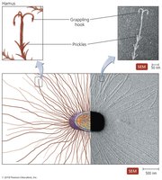
Archaeal Hami
Archaeal cells possess unique external appendages called hami, which are helical filaments with prickles and grappling hooks for surface attachment.
Transport Across Bacterial Cytoplasmic Membranes
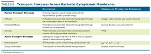
Transport Processes
Bacterial cells utilize various transport mechanisms to move substances across their cytoplasmic membranes.
Process | Description | Examples of Transported Substances |
|---|---|---|
Diffusion | Movement down electrochemical gradient through membrane | Oxygen, carbon dioxide, lipid-soluble chemicals |
Facilitated diffusion | Movement down electrochemical gradient through channels or carrier proteins | Glucose, fructose, urea, some vitamins |
Osmosis | Water molecules move down their concentration gradient | Water |
Active transport | ATP-dependent carrier proteins bring substances into cell | Na+, K+, Ca2+, H+, Cl- |
Group translocation | Substance is chemically altered during transport | Glucose, mannose, fructose |
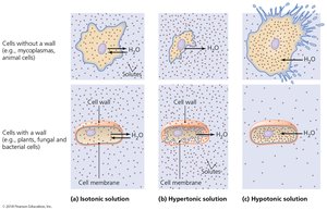
Effects of Solutions on Cells
The movement of water across cell membranes is influenced by the tonicity of the surrounding solution.
Isotonic: No net movement of water.
Hypertonic: Cells shrink due to water loss.
Hypotonic: Cells gain water; animal cells may burst, but cells with walls resist bursting.
Microbial Metabolism
ATP Production and Energy Storage
Cells produce ATP through three types of phosphorylation:
Substrate-level phosphorylation: Transfer of phosphate from an organic compound to ADP.
Oxidative phosphorylation: Energy from redox reactions is used to attach inorganic phosphate to ADP.
Photophosphorylation: Uses light energy to phosphorylate ADP.
Enzyme Activity and Regulation
Factors influencing enzyme activity: Temperature, pH, enzyme and substrate concentrations, presence of inhibitors.
Activators: Cofactors bind to allosteric sites to activate enzymes.
Inhibitors:
Competitive inhibitors: Bind to active site, blocking substrate.
Noncompetitive inhibitors: Bind to allosteric site, altering enzyme shape.
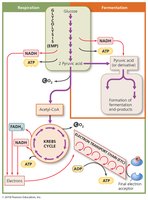
Carbohydrate Catabolism
Glycolysis: Occurs in cytosol, splits glucose into 2 pyruvate, 2 ATP, and 2 NADH.
Cellular respiration: Complete oxidation of substrates, ATP production via redox reactions. Stages:
Synthesis of acetyl-CoA
Krebs cycle
Electron transport chain (most significant ATP production)
Fermentation: Partial oxidation of sugar, uses organic molecule as final electron acceptor, less efficient than respiration.
Photosynthesis and Anabolic Pathways
Photosynthesis: Light energy is used to synthesize carbohydrates from CO2 and H2O.
Amphibolic reactions: Metabolic reactions that are reversible, functioning in both catabolic and anabolic pathways.
Gluconeogenesis: Synthesis of glucose from amino acids, glycerol, and fatty acids.
Chemiosmosis
Chemiosmosis: Ions flow down their electrochemical gradient across a membrane to synthesize ATP.
Summary Table: Key Microbial Groups
Group | Cell Type | Key Features |
|---|---|---|
Bacteria | Prokaryotic | Peptidoglycan cell wall, asexual reproduction |
Archaea | Prokaryotic | No peptidoglycan, extreme environments |
Fungi | Eukaryotic | Obtain food from others, cell wall, molds and yeasts |
Protozoa | Eukaryotic | Motile, single-celled, animal-like |
Algae | Eukaryotic | Photosynthetic, oxygen production |
Viruses | Acellular | Obligate parasites, protein coat |
Additional info: Academic context was added to clarify and expand brief points, ensuring completeness and self-contained explanations for exam preparation.
