 Back
BackMicrobiology Exam 1 Study Guide: History, Cell Structure, Microscopy, and Bacterial Growth
Study Guide - Smart Notes
 Tailored notes based on your materials, expanded with key definitions, examples, and context.
History of Microbiology
Pioneers and Their Contributions
The field of microbiology was shaped by several key scientists whose discoveries laid the foundation for our understanding of microorganisms and infectious diseases.
Anton van Leeuwenhoek: First to observe and describe microorganisms using a simple microscope.
Louis Pasteur: Disproved spontaneous generation, developed pasteurization, and contributed to the germ theory of disease.
Joseph Lister: Introduced antiseptic techniques in surgery, reducing infections.
Ignaz Semmelweis: Advocated handwashing to prevent puerperal fever in hospitals.
Robert Koch: Developed Koch’s postulates, linking specific microbes to specific diseases.
Edward Jenner: Developed the first successful smallpox vaccine.
Paul Ehrlich: Pioneered chemotherapy and discovered the first effective treatment for syphilis.
Alexander Fleming: Discovered penicillin, the first antibiotic.
J. Craig Venter: Sequenced many microbial genomes, advancing genomics.
Koch’s Postulates and Germ Theory
Koch’s Postulates: Criteria to establish a causative relationship between a microbe and a disease.
Relation to Germ Theory: Provided experimental evidence that specific microbes cause specific diseases.
Disproving Spontaneous Generation
Crucial Experiment: Louis Pasteur’s swan-neck flask experiment showed that microorganisms do not arise spontaneously but from other microbes.
Cell Structure and Function
What are the different possible shapes of bacteria and what are they called?
Cocci: Spherical
Bacilli: Rod-shaped
Spirilla: Spiral
Vibrios: Comma-shaped
Spirochetes: Flexible spirals
What do arrangement prefixes “strepto-” and “staphylo-” stand for?
Strepto-: Chains
Staphylo-: Clusters
What is the nature of the bacterial chromosome?
Typically a single, circular DNA molecule
What are the components in a bacterial cell wall? How does Gram+ cell walls differ from Gram– cell walls?
Main component: Peptidoglycan
Gram-positive: Thick peptidoglycan layer
Gram-negative: Thin peptidoglycan layer and an outer membrane containing lipopolysaccharide (LPS)
Why is LPS considered an endotoxin?
LPS can trigger strong immune responses and is toxic to animals
What is an endospore? What are endospores resistant to?
Highly resistant structures formed by some bacteria to survive harsh conditions
Resistant to heat, chemicals, desiccation, and radiation
Why do conjugation pili in certain microbes alarm health care professionals?
They facilitate DNA transfer between bacteria, spreading antibiotic resistance
What is the endosymbiotic hypothesis? What does it have to do with microbiology?
Suggests mitochondria and chloroplasts originated from engulfed prokaryotes
Explains the evolutionary origin of key eukaryotic organelles
How does facilitated diffusion differ from active transport?
Facilitated diffusion: Passive, moves substances down their concentration gradient via transport proteins
Active transport: Requires energy, moves substances against their concentration gradient via transport proteins
How does phagocytosis allow eukaryotic macrophages to get rid of bacteria?
Macrophages engulf bacteria into vesicles, which fuse with lysosomes for digestion and destruction
How does a bacterial (prokaryotic) flagellum differ from a eukaryotic one?
Bacterial flagella: Simpler, rotate for movement
Eukaryotic flagella: Complex, whip-like motion
How does a glycocalyx help a bacteria to be pathogenic?
Helps bacteria evade immune responses and adhere to surfaces
Why are bacteria able to endure some osmotic stress?
Bacterial cell walls provide protection against osmotic lysis
Do bacteria have different ribosomes than eukaryotic cells?
Yes, bacterial ribosomes are 70S; eukaryotic ribosomes are 80S
Can you identify the Eukaryotic organelles and their function?
Nucleus: Contains DNA
Mitochondria: Energy production
Endoplasmic reticulum: Protein and lipid synthesis
Golgi apparatus: Modifies and packages proteins/lipids
Lysosomes: Digestion
Chloroplasts (plants/algae): Photosynthesis
Others: Peroxisomes, vacuoles, cytoskeleton, ribosomes
Bacterial Shapes and Arrangements
Bacteria exhibit various shapes and arrangements, which are important for identification and classification.
Shapes: Cocci (spherical), Bacilli (rod-shaped), Spirilla (spiral), Vibrios (comma-shaped), Spirochetes (flexible spirals).
Arrangements: Strepto- (chains), Staphylo- (clusters).
Bacterial Cell Wall and Chromosome
Bacterial Chromosome: Typically a single, circular DNA molecule.
Cell Wall Components: Peptidoglycan is the main component. Gram-positive bacteria have thick peptidoglycan layers; Gram-negative bacteria have thin layers and an outer membrane containing lipopolysaccharide (LPS).
LPS as Endotoxin: LPS can trigger strong immune responses and is toxic to animals.
Endospores and Pili
Endospores: Highly resistant structures formed by some bacteria to survive harsh conditions.
Conjugation Pili: Facilitate DNA transfer between bacteria; important in the spread of antibiotic resistance.
Eukaryotic vs. Prokaryotic Features
Endosymbiotic Hypothesis: Suggests mitochondria and chloroplasts originated from engulfed prokaryotes.
Flagella: Bacterial flagella are simpler and rotate; eukaryotic flagella are complex and whip-like.
Glycocalyx: Helps bacteria evade immune responses and adhere to surfaces.
Osmotic Stress: Bacterial cell walls provide protection against osmotic lysis.
Ribosomes: Bacterial (70S) ribosomes differ from eukaryotic (80S) ribosomes.
Eukaryotic Organelles: Nucleus, mitochondria, endoplasmic reticulum, Golgi apparatus, lysosomes, etc.
Microscopy, Staining, and Classification
How are the properties of light exploited to visualize minute organisms?
Microscopy uses refraction—the bending of light as it passes through different media—to magnify and resolve tiny organisms.
What is the unit used to describe the wavelength of visible light?
Nanometer (nm); 1 nm = 10-9 meter.
The shorter the wavelength, the more energetic the light, and more likely to bend (blue).
Shorter wavelengths (like blue light) provide better resolution because they are more energetic and bend more.
Define the terms:
Refraction: Bending of light as it passes through different media.
Diffraction: Spreading of light waves as they pass around edges.
Absorption: Uptake of light energy by a material.
Reflection: Bouncing of light off a surface.
Transmission: Passage of light through a material.
Fluorescence: Emission of light by a substance that has absorbed light.
How can the contrast between a sample field and a specimen be increased?
By staining the specimen or using special microscopes (like phase contrast or DIC) to enhance differences between the specimen and background.
Why can an electron microscope give better resolution?
Electron microscopes use electrons, which have much shorter wavelengths than visible light, allowing for much higher resolution.
Why wouldn’t red light be a good choice for a microscope?
Red light has a longer wavelength, resulting in lower resolution compared to blue light.
Resolution Equation:
D = λ / NA Where D = minimal distance to distinguish objects, λ = wavelength, NA = numerical aperture.
Why can phase contrast or DIC microscopes work without stains?
They enhance contrast by exploiting differences in refractive index within the specimen, allowing visualization of live, unstained cells.
What is the advantage of confocal microscopy?
Confocal microscopy allows for 3D imaging and improved resolution by focusing on a single plane within the specimen.
What is a Gram stain?
A differential stain that classifies bacteria as Gram-positive or Gram-negative based on cell wall structure.
What are some of the tools that can help us to classify microorganisms?
Morphology, staining, biochemical tests, and genetic analysis.
What are the three Domains of life on earth?
Bacteria, Archaea, Eukarya
What is the biological binomial nomenclature for a Human?
Homo sapiens
Principles of Microscopy
Microscopy is essential for visualizing microorganisms, exploiting the properties of light to achieve magnification and resolution.
Refraction: Bending of light as it passes through different media.
Diffraction: Spreading of light waves as they pass around edges.
Absorption: Uptake of light energy by a material.
Reflection: Bouncing of light off a surface.
Transmission: Passage of light through a material.
Fluorescence: Emission of light by a substance that has absorbed light.
Improving Contrast and Resolution
Contrast: Enhanced by staining or using special microscopes (phase contrast, DIC).
Electron Microscopy: Provides higher resolution due to shorter electron wavelengths.
Wavelength: Shorter wavelengths (e.g., blue light) provide better resolution; red light is less effective.
Resolution Equation:
Where D = minimal distance to distinguish objects, \( \lambda \) = wavelength, NA = numerical aperture.
Confocal Microscopy: Allows for 3D imaging and improved resolution.
Staining and Classification
Gram Stain: Differentiates bacteria into Gram-positive and Gram-negative based on cell wall structure.
Classification Tools: Morphology, staining, biochemical tests, genetic analysis.
Three Domains of Life: Bacteria, Archaea, Eukarya.
Binomial Nomenclature for Humans: Homo sapiens.
Nutrition and Growth of Bacteria
🦠 Bacteria require:
Sources of carbon, nitrogen, energy, and electrons to grow.
🌱 Four classes of bacteria based on their sources:
Photoautotrophs: Energy from light, carbon from CO2 in air.
Chemoautotrophs: Energy from chemicals, carbon from CO2 in air.
Photoheterotrophs: Energy from light, carbon from organic molecules.
Chemoheterotrophs: Energy and carbon from organic molecules (usually sugars). Most lab bacteria are in this group.
🌬️ Oxygen requirements:
Aerobic: Require oxygen for cellular respiration.
Anaerobic: Do not require oxygen.
Four classes: Obligate aerobes, Obligate anaerobes, Facultative anaerobes, Aerotolerant anaerobes (can be determined with FTM tubes).
🌡️ Temperature requirements:
Mesophiles: Prefer moderate temperatures (studied in lab).
Psychrophiles: Prefer cold.
Thermophiles: Prefer high temperatures.
🧪 pH requirements:
Acidophiles: Like low pH.
Neutrophiles: Prefer pH 7.
💧 Water & Osmotic Balance:
Bacteria need water to grow and depend on proper osmotic balance.
👥 Quorum Sensing:
Some bacteria sense and respond to the presence of other bacteria, preferring to grow in crowds.
🍲 Culture Media Types:
Rich, defined: All nutrients in known ratios.
Minimal defined: Only essential nutrients in known quantities.
Rich undefined: Complex nutrients, not all quantified (e.g., TSA media).
Selective media: Only certain organisms grow (e.g., MacConkey’s agar for Gram negatives).
Differential media: Color indicator distinguishes organisms (e.g., MacConkey’s agar turns colonies red if lactose is used).
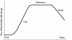
📈 Growth Phases:
Lag phase: Slow growth.
Log phase: Fast growth (most metabolically active).
Stationary phase: No growth.
Decline phase: Death.
🦠 Bacterial Growth:
Occurs by binary fission (each cell splits into two).
Growth is geometric (logarithmic).
Doubling time: Time for one division (e.g., Vibrio harveyii = 10 min).
Chemostat: Special incubator keeps bacteria in log phase by adding media and removing waste.
🔍 Measuring Bacterial Populations:
Direct microscopy and counting grid slides.
Turbidity measurements.
Plating after serial dilution for direct colony forming unit counting.
Requirements for Bacterial Growth
Bacteria require sources of carbon, nitrogen, energy, and electrons for growth. They are classified based on how they obtain these resources.
Photoautotrophs: Use light for energy and CO2 for carbon.
Chemoautotrophs: Use chemicals for energy and CO2 for carbon.
Photoheterotrophs: Use light for energy and organic molecules for carbon.
Chemoheterotrophs: Use chemicals and organic molecules for both energy and carbon (most lab bacteria).
Oxygen Requirements
Obligate Aerobes: Require oxygen.
Obligate Anaerobes: Cannot tolerate oxygen.
Facultative Anaerobes: Can grow with or without oxygen.
Aerotolerant Anaerobes: Tolerate oxygen but do not use it.
Physical Requirements for Growth
Temperature: Mesophiles (moderate), psychrophiles (cold), thermophiles (hot).
pH: Acidophiles (low pH), neutrophiles (neutral pH).
Water: Essential for all bacteria; osmotic balance is critical.
Quorum Sensing
Bacteria can sense population density and coordinate behavior in groups.
Types of Media
Type of Media | Description | Example |
|---|---|---|
Rich, Defined | All nutrients in known ratios | Minimal defined media |
Rich, Undefined | Complex nutrients, not all quantified | TSA media |
Selective | Allows only certain organisms to grow | MacConkey’s agar (Gram-negatives) |
Differential | Distinguishes organisms by color change | MacConkey’s agar (lactose fermenters turn red) |
Bacterial Growth Curve
Bacterial populations grow in distinct phases: lag, log (exponential), stationary, and death. Growth is geometric (logarithmic) due to binary fission. The time for one division is the doubling time.
Lag Phase: Adaptation, little growth.
Log Phase: Rapid, exponential growth; cells are most metabolically active.
Stationary Phase: Growth rate slows; nutrients deplete, waste accumulates.
Death Phase: Cells die due to lack of nutrients and accumulation of toxins.
Chemostat: Device that maintains bacterial cultures in log phase by continuous nutrient supply and waste removal.
Measuring Growth: Direct counts (microscopy), turbidity, and colony-forming unit (CFU) counts after serial dilution.
