 Back
Microbiology Foundations: The Microbial World, Cell Structure, and Growth
Study Guide - Smart Notes
 Tailored notes based on your materials, expanded with key definitions, examples, and context.
The Microbial World and You
Introduction to Microorganisms
Microorganisms, or microbes, are minute living entities that are typically too small to be seen without a microscope. They play essential roles in ecosystems, human health, and disease. The major groups of microorganisms include bacteria, archaea, fungi, protozoa, microscopic algae, viruses, and helminths (parasitic worms).
Bacteria: Unicellular prokaryotes with diverse shapes (bacillus, coccus, spiral) and arrangements (chains, clusters). Cell walls contain peptidoglycan. Reproduce by binary fission.
Archaea: Prokaryotes lacking peptidoglycan in their cell walls. Often found in extreme environments (e.g., halophiles, thermophiles).
Fungi: Eukaryotes with chitin cell walls. Can be unicellular (yeasts) or multicellular (molds, mushrooms).
Protozoa: Unicellular eukaryotes with various modes of locomotion (pseudopods, flagella, cilia). Some are parasitic.
Algae: Photosynthetic eukaryotes with cellulose cell walls. Produce oxygen and organic compounds.
Viruses: Acellular entities with a core of DNA or RNA surrounded by a protein coat. Replicate only inside host cells.
Helminths: Multicellular animal parasites (flatworms, roundworms) with microscopic life stages.
Classification of Organisms
Organisms are classified into three domains based on cellular and molecular characteristics:
Bacteria: Prokaryotes with peptidoglycan cell walls.
Archaea: Prokaryotes with cell walls lacking peptidoglycan.
Eukarya: Eukaryotes, including protists, fungi, plants, and animals.
Spontaneous Generation vs. Biogenesis
Historically, the origin of life was debated between spontaneous generation (life arises from nonliving matter) and biogenesis (life arises from preexisting life). Key experiments disproved spontaneous generation and established the principle of biogenesis.
Francesco Redi (1668): Demonstrated that maggots do not arise spontaneously from decaying meat.
John Needham (1745): Supported spontaneous generation with boiled broth experiments.
Lazzaro Spallanzani (1765): Showed that boiling and sealing broth prevented microbial growth, supporting biogenesis.
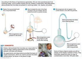
Louis Pasteur (1861): Used swan-neck flasks to show that microorganisms are present in the air and do not arise spontaneously from nonliving matter.
The Golden Age of Microbiology
Major Discoveries and Contributors (1857–1914)
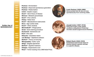
This era saw foundational discoveries linking microbes to fermentation, disease, and immunity, and the development of aseptic techniques and antimicrobial therapies.
Louis Pasteur: Disproved spontaneous generation, developed pasteurization, and established the role of microbes in fermentation and disease.
Joseph Lister: Introduced antiseptic surgery using phenol, reducing surgical infections.
Robert Koch: Developed Koch's postulates, linking specific microbes to specific diseases (e.g., anthrax, tuberculosis).
Edward Jenner: Developed the smallpox vaccine using cowpox virus.
Discovery of antibiotics: Alexander Fleming discovered penicillin, the first true antibiotic.
Functional Anatomy of Prokaryotic Cells
Overview of Bacterial Cell Structure
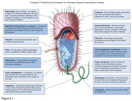
Bacterial cells possess unique structures that contribute to their survival, pathogenicity, and classification. Key features include the cell wall, plasma membrane, nucleoid, plasmids, ribosomes, and external appendages.
Peptidoglycan Structure
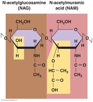
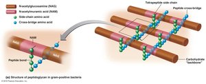
The bacterial cell wall is primarily composed of peptidoglycan, a macromolecular network unique to bacteria. Peptidoglycan consists of repeating disaccharide units of N-acetylglucosamine (NAG) and N-acetylmuramic acid (NAM), cross-linked by short peptides.
NAG and NAM: The glycan backbone is formed by alternating NAG and NAM residues.
Tetrapeptide side chains: Attached to NAM, these peptides are cross-linked to provide structural strength.
Gram-Positive vs. Gram-Negative Cell Walls
Bacteria are classified as Gram-positive or Gram-negative based on cell wall structure and staining properties:
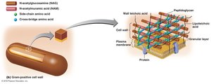
Gram-Positive: Thick peptidoglycan layer, teichoic acids, no outer membrane. Retain crystal violet stain (purple).
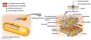
Gram-Negative: Thin peptidoglycan layer, outer membrane containing lipopolysaccharide (LPS), periplasmic space. Do not retain crystal violet; counterstained pink/red.
Lipopolysaccharide (LPS) Components
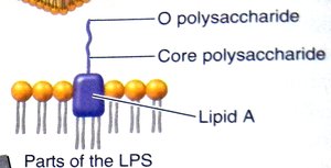
LPS is a major component of the outer membrane of Gram-negative bacteria and consists of:
Lipid A: Endotoxin responsible for fever and shock.
Core polysaccharide: Provides stability.
O polysaccharide: Functions as an antigen for identification.
Gram Staining Technique
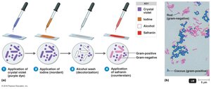
The Gram stain is a differential staining method that distinguishes bacteria based on cell wall structure:
Application of crystal violet (primary stain)
Addition of iodine (mordant)
Alcohol wash (decolorization)
Application of safranin (counterstain)
Gram-positive bacteria remain purple; Gram-negative bacteria appear pink/red.
Microbial Growth
Physical and Chemical Requirements for Growth
Microbial growth depends on various physical and chemical factors:
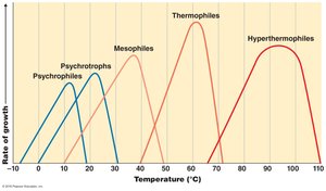
Temperature: Microbes are classified by their optimal growth temperatures: psychrophiles (cold-loving), mesophiles (moderate), thermophiles (heat-loving), and hyperthermophiles (extreme heat).
pH: Most bacteria grow best near neutral pH; acidophiles and alkaliphiles thrive in acidic or basic environments, respectively.
Osmotic Pressure: Halophiles require or tolerate high salt concentrations; plasmolysis occurs in hypertonic environments.
Bacterial Growth Curve
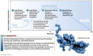
Bacterial populations exhibit characteristic growth phases in batch culture:
Lag phase: Adaptation, no increase in cell number.
Log (exponential) phase: Rapid cell division and population growth.
Stationary phase: Growth rate slows as resources become limited.
Death phase: Cells die at an exponential rate.
Classification of Microorganisms
The Three-Domain System
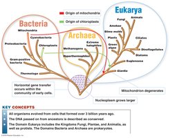
Microorganisms are classified into three domains based on genetic and biochemical differences: Bacteria, Archaea, and Eukarya. This system reflects evolutionary relationships and the origin of eukaryotic organelles via endosymbiosis.
Summary Table: Key Differences Between Gram-Positive and Gram-Negative Bacteria
Feature | Gram-Positive | Gram-Negative |
|---|---|---|
Peptidoglycan Layer | Thick | Thin |
Teichoic Acids | Present | Absent |
Outer Membrane | Absent | Present (contains LPS) |
Stain Color | Purple | Pink/Red |
Sensitivity to Penicillin | High | Low |
Additional info: This guide covers foundational concepts from the first chapters of a college-level microbiology course, including microbial diversity, cell structure, classification, and growth. It is suitable for exam preparation and as a reference for laboratory techniques and microbial physiology.
