 Back
BackMicrobiology Lab Exam 1 Study Guide: Microscopy, Staining, Media, and Lab Techniques
Study Guide - Smart Notes
 Tailored notes based on your materials, expanded with key definitions, examples, and context.
Microscopy and Staining Techniques
Types of Stains and Their Applications
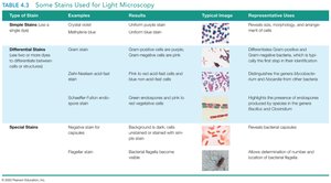
Staining is a fundamental technique in microbiology that enhances the contrast of microscopic specimens, allowing for the visualization and differentiation of microbial cells and their structures. Stains can be classified as simple, differential, or special, each serving distinct purposes in microbial identification and study.
Simple Stains: Use a single dye (e.g., crystal violet, methylene blue) to color all cells uniformly, revealing cell shape, morphology, and arrangement.
Differential Stains: Employ multiple dyes to distinguish between different types of bacteria or cellular components. Examples include the Gram stain (differentiates Gram-positive and Gram-negative bacteria), acid-fast stain (identifies acid-fast bacteria like Mycobacterium), and endospore stain (highlights bacterial endospores).
Special Stains: Target specific structures such as capsules (capsule stain) or flagella (flagella stain), aiding in the identification of unique bacterial features.
Key Points:
Simple stains provide basic information about cell shape and arrangement.
Gram stain is crucial for bacterial classification and guides clinical treatment decisions.
Acid-fast stain is used for identifying pathogens like Mycobacterium tuberculosis.
Endospore and capsule stains reveal structures important for bacterial survival and pathogenicity.
Example: A Gram stain showing purple (Gram-positive) cocci in clusters suggests Staphylococcus species, while pink (Gram-negative) rods may indicate Escherichia coli.
Laboratory Equipment and Media
Common Lab Tools and Their Functions
Microbiology labs utilize specialized equipment to culture, observe, and manipulate microorganisms safely and effectively. Understanding the function of each tool is essential for proper technique and safety.
Inoculating loop/needle: Used to transfer microorganisms to media.
Bacticinerator: Sterilizes inoculating tools using heat.
Autoclave: Sterilizes media and equipment using pressurized steam.
Bibulous paper: Used for blotting slides after staining.
Optical lens wipe: Cleans microscope lenses without scratching.
Types of Culture Media
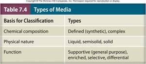
Culture media provide the nutrients required for microbial growth and can be classified based on chemical composition, physical state, and function. The choice of medium affects the ability to isolate, identify, and study microorganisms.
Basis for Classification | Types |
|---|---|
Chemical composition | Defined (synthetic), complex |
Physical nature | Liquid, semisolid, solid |
Function | Supportive (general purpose), enriched, selective, differential |
Defined media: Exact chemical composition is known.
Complex media: Contains ingredients of unknown composition (e.g., nutrient agar).
Selective media: Inhibits growth of some organisms while allowing others (e.g., EMB, MSA).
Differential media: Distinguishes organisms based on metabolic reactions (e.g., color change on MSA or EMB).
Supportive media: Supports the growth of many organisms without selection or differentiation.
Example: Eosin methylene blue (EMB) agar is both selective (inhibits Gram-positive bacteria) and differential (identifies lactose fermenters by color change).
Microbial Identification and Lab Techniques
Streak Plate Method and Colony Isolation
The streak plate technique is used to isolate pure colonies from a mixed culture by spreading bacteria over the surface of an agar plate in a pattern that thins out the sample and separates individual cells.
Purpose: To obtain isolated colonies for further study.
Procedure: Involves streaking the inoculum across the plate in sections (three, four, or five), sterilizing the loop between sections.
Correct orientation: Plates are incubated upside down to prevent condensation from dripping onto the agar surface.
Viable Plate Count and Dilution Calculations
Viable plate count is a quantitative method to estimate the number of living bacteria in a sample. It involves serial dilution and plating, followed by colony counting.
Equation for CFU/ml:
Countable plate range: 30–300 CFU.
Pour plate: Uses 1 ml inoculum; Spread plate: Uses 0.1 ml inoculum.
Example: If 100 colonies are counted on a plate inoculated with 0.1 ml of a 10-4 dilution, the original concentration is:
Lab Safety and Aseptic Technique
Safety Procedures and Biosafety Levels
Proper lab safety prevents the escape of microorganisms and protects both the experimenter and the environment. Biosafety levels (BSL) classify organisms based on risk:
BSL-1: Non-pathogenic organisms; basic safety.
BSL-2: Moderate risk; lab coats, gloves, and limited access required.
BSL-3: Pathogens with potential for aerosol transmission; special ventilation.
BSL-4: High-risk, life-threatening agents; maximum containment.
Example: Most teaching labs, including Three Rivers, use BSL-1 or BSL-2 organisms.
Aseptic Technique
Aseptic technique is essential to prevent contamination of cultures and the environment. It involves sterilizing tools, minimizing exposure, and using proper transfer methods.
Goals: Prevent contamination of the environment and the culture.
Steps: Sterilize inoculating loop, avoid touching non-sterile surfaces, flame tube openings, and work near a flame or bacticinerator.
Microscope Use and Care
Parts and Functions of the Microscope
Understanding the microscope's components and their functions is crucial for effective observation of microorganisms.
Objective lenses: 4x (scanning), 10x (low power), 40x (high dry), 100x (oil immersion).
Total magnification: Calculated by multiplying the ocular lens (usually 10x) by the objective lens.
Parfocal lenses: Remain in focus when switching between objectives.
Proper use: Start with the lowest power, use coarse then fine focus, and adjust light intensity as needed.
Key Microbial Species and Their Laboratory Identification
Representative Bacteria and Their Features
Familiarity with common lab organisms and their distinguishing features is essential for identification in practical exams.
Bacillus subtilis: Gram-positive rods, short chains, forms endospores.
Escherichia coli: Gram-negative coccobacilli, metallic green on EMB, coliform.
Micrococcus luteus: Gram-positive cocci, tetrads.
Mycobacterium tuberculosis: Acid-fast rod.
Staphylococcus epidermidis: Gram-positive cocci, clusters, pink/red on MSA.
Staphylococcus aureus: Gram-positive cocci, clusters, yellow on MSA.
Serratia marcescens: Gram-negative rods, red colonies at 30°C.
Enterobacter aerogenes: Gram-negative rods, pigmented on EMB, coliform.
Alcaligenes faecalis: Gram-negative rod, colorless on EMB, non-coliform.
Example: Identification of Staphylococcus aureus is based on Gram-positive cocci in clusters and yellow colonies on MSA.
Terminology Clarification
Commonly Confused Terms
Cell: Single microbial unit.
Colony: Visible mass of cells derived from a single progenitor.
Culture: Growth of microorganisms in or on a medium.
Smear: Thin layer of bacteria on a slide for staining.
Streak: Technique for isolating colonies on a plate.
Spread: Even distribution of inoculum over the surface of a plate.
Additional info: Mastery of these terms is essential for accurate communication and documentation in laboratory settings.
