 Back
BackMicrobiology Lab Final Exam Review – Step-by-Step Guidance
Study Guide - Smart Notes
 Tailored notes based on your materials, expanded with key definitions, examples, and context.
Q1. What is the purpose of a White Blood Count (WBC)?
Background
Topic: Hematology – White Blood Cells
This question tests your understanding of why WBC counts are performed in clinical settings and what information they provide about a patient's health.
Key Terms:
White Blood Cells (WBC): Cells of the immune system involved in protecting the body against infections.
White Blood Count: A laboratory test that measures the number of WBCs in blood.
Step-by-Step Guidance
Consider the role of WBCs in the immune response and how their levels can indicate infection, inflammation, or other health conditions.
Think about how a WBC count is used diagnostically to assess overall health or detect abnormalities.
Reflect on situations where a WBC count might be ordered (e.g., suspected infection, monitoring immune status).
Try solving on your own before revealing the answer!
Final Answer:
The purpose of a White Blood Count (WBC) is to measure the number of white blood cells in the blood, which helps assess the body's immune status and detect conditions such as infection, inflammation, immune disorders, or hematologic diseases.
Q2. Define leukocytosis, leukopenia, neutrophilic leukocytosis, eosinophilia, basophilia, monocytosis, and lymphocytosis.
Background
Topic: Hematology – White Blood Cell Disorders
This question tests your knowledge of terminology related to abnormal WBC counts and specific cell type increases or decreases.
Key Terms:
Leukocytosis: Increased total WBC count.
Leukopenia: Decreased total WBC count.
Neutrophilic leukocytosis: Increased neutrophils.
Eosinophilia: Increased eosinophils.
Basophilia: Increased basophils.
Monocytosis: Increased monocytes.
Lymphocytosis: Increased lymphocytes.
Step-by-Step Guidance
Recall the normal ranges for WBCs and their subtypes.
Think about what it means when a cell type is "-cytosis" (increase) or "-penia" (decrease).
Match each term to the specific cell type and whether it refers to an increase or decrease.
Try solving on your own before revealing the answer!
Final Answer:
Leukocytosis: Increased total WBC count.
Leukopenia: Decreased total WBC count.
Neutrophilic leukocytosis: Increased neutrophils.
Eosinophilia: Increased eosinophils.
Basophilia: Increased basophils.
Monocytosis: Increased monocytes.
Lymphocytosis: Increased lymphocytes.
Q3. Identify white blood cells in a blood smear.
Background
Topic: Microscopy – Blood Smear Analysis
This question tests your ability to recognize different types of WBCs under the microscope based on their morphology.
Key Terms:
Blood Smear: A thin layer of blood spread on a microscope slide.
White Blood Cells: Includes neutrophils, lymphocytes, monocytes, eosinophils, and basophils.
Step-by-Step Guidance
Review the distinguishing features of each WBC type (e.g., nucleus shape, cytoplasmic granules).
Compare cell size, nuclear morphology, and granule presence to identify each cell.
Practice matching cell types to their visual characteristics in a blood smear.
Try solving on your own before revealing the answer!
Final Answer:
White blood cells in a blood smear can be identified by their size, nuclear shape, and granules: neutrophils (multi-lobed nucleus), lymphocytes (large round nucleus), monocytes (kidney-shaped nucleus), eosinophils (bilobed nucleus, red granules), basophils (bilobed nucleus, blue granules).
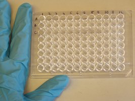
Q4. What is pictured in the image below?
Background
Topic: Immunology – ELISA Test
This question tests your ability to recognize laboratory equipment used in immunological assays.
Key Terms:
ELISA Plate: A 96-well plate used for enzyme-linked immunosorbent assay.
Step-by-Step Guidance
Observe the structure and layout of the plate (rows and columns of wells).
Recall common uses for this type of plate in immunology labs.
Think about the types of tests that require multiple samples or controls in a grid format.
Try solving on your own before revealing the answer!
Final Answer:
The image shows a 96-well ELISA plate, commonly used for enzyme-linked immunosorbent assays to detect antigens or antibodies in samples.
Q5. List the steps of an ELISA test.
Background
Topic: Immunology – ELISA Technique
This question tests your understanding of the procedure for performing an ELISA (enzyme-linked immunosorbent assay).
Key Terms:
ELISA: A plate-based assay technique for detecting and quantifying substances such as peptides, proteins, antibodies, and hormones.
Step-by-Step Guidance
Recall the sequence of steps: coating, blocking, sample addition, detection antibody addition, substrate addition.
Think about the purpose of each step (e.g., blocking prevents nonspecific binding).
Consider how the final color change indicates a positive result.
Try solving on your own before revealing the answer!
Final Answer:
Coat wells with antigen or antibody.
Block nonspecific binding sites.
Add sample (containing target analyte).
Add detection antibody.
Add enzyme-linked secondary antibody.
Add substrate; observe color change.
Each step is designed to ensure specificity and sensitivity in detecting the target molecule.
Q6. List a step-by-step summary of the order things bind to each other in a positive well of an ELISA test.
Background
Topic: Immunology – ELISA Binding Sequence
This question tests your understanding of the molecular interactions in a positive ELISA well.
Key Terms:
Antigen, Antibody, Enzyme, Substrate
Step-by-Step Guidance
Recall the initial binding of antigen or antibody to the well.
Think about the sequence of additions: sample, detection antibody, enzyme-linked antibody, substrate.
Consider how each binding step leads to the final detectable signal.
Try solving on your own before revealing the answer!
Final Answer:
Antigen (or antibody) binds to the well.
Sample antibody (or antigen) binds to the coated antigen (or antibody).
Enzyme-linked secondary antibody binds to the sample antibody.
Substrate binds to the enzyme, producing a color change.
Q7. Be able to identify a positive sample in an ELISA test.
Background
Topic: Immunology – ELISA Interpretation
This question tests your ability to interpret ELISA results based on color change or optical density.
Key Terms:
Positive Sample: Indicates presence of target analyte.
Color Change: Usually yellow or blue, depending on substrate.
Step-by-Step Guidance
Recall what a positive result looks like (color change in the well).
Compare wells with and without color change.
Think about how controls are used to validate the result.
Try solving on your own before revealing the answer!
Final Answer:
A positive sample in an ELISA test is identified by a color change in the well, indicating the presence of the target antigen or antibody.
Q8. How can PCR be used in the lab? What can it tell us?
Background
Topic: Molecular Biology – PCR Applications
This question tests your understanding of the uses and informational value of polymerase chain reaction (PCR) in laboratory settings.
Key Terms:
PCR: Polymerase Chain Reaction, a technique to amplify DNA.
Step-by-Step Guidance
Recall the main purpose of PCR: DNA amplification.
Think about how PCR can be used to detect pathogens, genetic mutations, or for cloning.
Consider the types of information PCR results provide (presence/absence, quantity, sequence).
Try solving on your own before revealing the answer!
Final Answer:
PCR can be used to amplify specific DNA sequences, allowing detection of pathogens, identification of genetic mutations, and analysis of genetic material. It tells us whether a target DNA is present and can quantify its amount.
Q9. What are the ingredients of PCR and the function of each ingredient?
Background
Topic: Molecular Biology – PCR Components
This question tests your knowledge of the essential reagents required for PCR and their roles.
Key Terms:
DNA Template, Primers, DNA Polymerase, dNTPs, Buffer
Step-by-Step Guidance
List the main ingredients used in PCR.
Describe the function of each ingredient (e.g., primers provide specificity).
Think about how each component contributes to successful DNA amplification.
Try solving on your own before revealing the answer!
Final Answer:
DNA Template: The sequence to be amplified.
Primers: Short DNA sequences that initiate replication.
DNA Polymerase: Enzyme that synthesizes new DNA.
dNTPs: Nucleotide building blocks for new DNA.
Buffer: Maintains optimal conditions for the reaction.
Q10. What are the steps of PCR?
Background
Topic: Molecular Biology – PCR Process
This question tests your understanding of the thermal cycling steps involved in PCR.
Key Terms:
Denaturation, Annealing, Extension
Step-by-Step Guidance
Recall the three main steps: denaturation, annealing, extension.
Think about the temperature and purpose of each step.
Consider how these steps are repeated for multiple cycles to amplify DNA.
Try solving on your own before revealing the answer!
Final Answer:
Denaturation: DNA strands are separated by heating.
Annealing: Primers bind to the target sequence.
Extension: DNA polymerase synthesizes new DNA.
These steps are repeated for 20–40 cycles to amplify the target DNA.
Q11. Define antimicrobials and zone of inhibition.
Background
Topic: Microbiology – Antimicrobial Testing
This question tests your understanding of terms related to antibiotic effectiveness.
Key Terms:
Antimicrobials: Agents that kill or inhibit microorganisms.
Zone of Inhibition: Area around an antibiotic disk where bacteria do not grow.
Step-by-Step Guidance
Recall the definition of antimicrobials and their role in treating infections.
Think about how the zone of inhibition is measured in disk diffusion tests.
Consider what a larger or smaller zone indicates about antibiotic effectiveness.
Try solving on your own before revealing the answer!
Final Answer:
Antimicrobials are substances that kill or inhibit the growth of microorganisms. The zone of inhibition is the clear area around an antibiotic disk where bacteria cannot grow, indicating the effectiveness of the antibiotic.
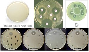
Q12. Describe the Kirby Bauer (disk diffusion) method.
Background
Topic: Microbiology – Antibiotic Susceptibility Testing
This question tests your knowledge of a standard method for evaluating antibiotic effectiveness.
Key Terms:
Kirby Bauer Method: Disk diffusion test for antibiotic susceptibility.
Mueller-Hinton Agar: Standard medium for this test.
Step-by-Step Guidance
Recall the steps: inoculate agar, place antibiotic disks, incubate, measure zones.
Think about how results are interpreted based on zone size.
Consider the importance of standardization (e.g., agar type, incubation time).
Try solving on your own before revealing the answer!
Final Answer:
The Kirby Bauer method involves inoculating a Mueller-Hinton agar plate with bacteria, placing antibiotic disks on the surface, incubating, and measuring the zones of inhibition to determine susceptibility.
Q13. If you are performing a Kirby Bauer test and antibiotic X has a zone of inhibition of 20-mm and antibiotic Y has a zone of inhibition of 40-mm, which one is more effective?
Background
Topic: Microbiology – Antibiotic Effectiveness
This question tests your ability to interpret disk diffusion results.
Key Terms:
Zone of Inhibition: Indicates effectiveness of antibiotic.
Step-by-Step Guidance
Recall what a larger zone of inhibition means in terms of bacterial susceptibility.
Compare the sizes of the zones for antibiotics X and Y.
Think about which antibiotic would be considered more effective based on the zone size.
Try solving on your own before revealing the answer!
Final Answer:
Antibiotic Y, with a 40-mm zone of inhibition, is more effective than antibiotic X, which has a 20-mm zone.
Q14. A Mueller-Hinton agar plate was inoculated with a lawn of Escherichia coli. Antibiotic (antimicrobial) disks were then placed on top of the agar media. Plates were incubated at 37°C for 24 hrs. The E. coli would be considered resistant to which antibiotic?
Background
Topic: Microbiology – Antibiotic Resistance
This question tests your ability to interpret disk diffusion results for resistance.
Key Terms:
Resistant: Bacteria grow up to the edge of the disk.
Zone of Inhibition: Indicates susceptibility.
Step-by-Step Guidance
Examine the plate for disks with little or no clear zone around them.
Recall that resistance is indicated by bacterial growth up to the disk.
Identify which disks show this pattern.
Try solving on your own before revealing the answer!
Final Answer:
E. coli would be considered resistant to antibiotics F and G, as there is little to no clear zone of inhibition and bacterial growth is up to the edge of the disks.
Q15. Define selective and differential media.
Background
Topic: Microbiology – Culture Media
This question tests your understanding of media types used to isolate and identify bacteria.
Key Terms:
Selective Media: Inhibits growth of some organisms, allows others.
Differential Media: Distinguishes organisms based on biochemical reactions.
Step-by-Step Guidance
Recall the purpose of selective media in isolating specific bacteria.
Think about how differential media helps identify bacteria based on color changes or reactions.
Consider examples of each type of media.
Try solving on your own before revealing the answer!
Final Answer:
Selective media inhibit the growth of certain bacteria while allowing others to grow. Differential media distinguish between organisms based on observable reactions, such as color changes.
Q16. Describe a positive and negative catalase reaction.
Background
Topic: Microbiology – Enzyme Tests
This question tests your ability to interpret catalase test results.
Key Terms:
Catalase: Enzyme that breaks down hydrogen peroxide.
Positive Reaction: Bubbling due to oxygen release.
Negative Reaction: No bubbling.
Step-by-Step Guidance
Recall the procedure: add hydrogen peroxide to bacterial colony.
Observe for bubbling (positive) or no reaction (negative).
Think about which bacteria are catalase positive or negative.
Try solving on your own before revealing the answer!
Final Answer:
A positive catalase reaction produces bubbles when hydrogen peroxide is added, indicating the presence of catalase. A negative reaction shows no bubbles.
Q17. Describe a positive and negative oxidase test.
Background
Topic: Microbiology – Enzyme Tests
This question tests your ability to interpret oxidase test results.
Key Terms:
Oxidase: Enzyme involved in electron transport chain.
Positive Reaction: Color change to purple/blue.
Negative Reaction: No color change.
Step-by-Step Guidance
Recall the procedure: add oxidase reagent to bacterial colony.
Observe for color change (positive) or no change (negative).
Think about which bacteria are oxidase positive or negative.
Try solving on your own before revealing the answer!
Final Answer:
A positive oxidase test results in a color change to purple or blue, indicating the presence of cytochrome oxidase. A negative test shows no color change.
Q18. State the purpose and be able to interpret results for the following media:
Background
Topic: Microbiology – Biochemical Media Interpretation
This question tests your knowledge of various media used to identify bacterial species and their metabolic capabilities.
Key Terms:
Starch plate, Urea agar, Milk agar, DNase agar, Gelatin tubes, Tributyrin agar plate, Blood agar plate, MacConkey agar, Eosin Methylene Blue agar, Hektoen agar, Citrate, Phenylalanine deaminase slant, Phenol red broth tubes, SIM media, MRVP, Nitrate reduction broth, Decarboxylase test, Motility test
Step-by-Step Guidance
Recall the purpose of each medium (e.g., starch plate tests for amylase activity).
Think about the expected positive and negative reactions for each medium.
Consider how color changes, clearing, or other observable results indicate metabolic activity.
Review examples of interpretation for each medium.
Try solving on your own before revealing the answer!
Final Answer:
Starch plate: Tests for amylase; clearing indicates positive.
Urea agar: Tests for urease; pink color is positive.
Milk agar: Tests for caseinase; clearing is positive.
DNase agar: Tests for DNase; clearing is positive.
Gelatin tubes: Tests for gelatinase; liquefaction is positive.
Tributyrin agar plate: Tests for lipase; clearing is positive.
Blood agar plate: Tests for hemolysis; clearing or color change is positive.
MacConkey agar: Selective/differential; pink colonies are positive for lactose fermentation.
Eosin Methylene Blue agar: Selective/differential; metallic green is positive for strong lactose fermenters.
Hektoen agar: Selective/differential; black precipitate is positive for H2S production.
Citrate: Tests for citrate utilization; blue color is positive.
Phenylalanine deaminase slant: Green color is positive.
Phenol red broth tubes: Yellow color is positive for fermentation.
SIM media: Tests for sulfur, indole, motility; black, red, and growth away from stab are positive.
MRVP: Red color is positive for MR; pink/red is positive for VP.
Nitrate reduction broth: Red color after reagents is positive.
Decarboxylase test: Purple color is positive.
Motility test: Growth away from stab is positive.
