 Back
Microbiology Lab Introduction Study Guide
Study Guide - Smart Notes
 Tailored notes based on your materials, expanded with key definitions, examples, and context.
Introduction to Microbiology Lab
Lab Safety and Preparation
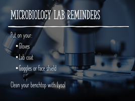
Microbiology labs require strict adherence to safety protocols to prevent contamination and ensure accurate results. Proper attire and workspace cleanliness are essential before beginning any experiment.
Personal Protective Equipment (PPE): Always wear gloves, a lab coat, and goggles or a face shield.
Workspace Preparation: Clean your benchtop with disinfectant (e.g., Lysol) before starting work.
Binomial Nomenclature and Taxonomy
Binomial Nomenclature
Binomial nomenclature is the formal system of naming species. Each organism is given a two-part scientific name: the genus and the species. This system, developed by Carl Linnaeus, ensures that each species has a unique and universally accepted name.
Genus: The first part of the name; always capitalized and italicized (or underlined).
Species: The second part; lowercase and italicized (or underlined).
Example: Escherichia coli (abbreviated as E. coli after first use).
Origin: Names are often derived from Latin or Greek, and may describe characteristics or honor scientists.
Correct Formatting: Genus species (e.g., Staphylococcus aureus).
Taxonomic Hierarchy
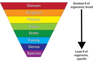
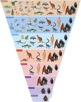
Taxonomy classifies organisms into a hierarchical structure, from the broadest category (domain) to the most specific (species). Each level is called a taxon.
Hierarchy (from broadest to most specific): Domain > Kingdom > Phylum > Class > Order > Family > Genus > Species
Each higher taxon includes more organisms but with less similarity.
Examples of Microbial Taxonomy
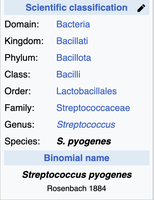
Streptococcus pyogenes: Domain: Bacteria, Kingdom: Bacillati, Phylum: Bacillota, Class: Bacilli, Order: Lactobacillales, Family: Streptococcaceae, Genus: Streptococcus, Species: S. pyogenes
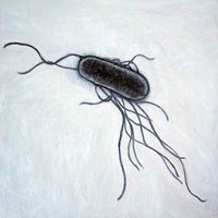
Escherichia coli: Genus honors Theodor Escherich; species describes its habitat (colon).
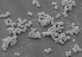
Staphylococcus aureus: Genus describes clustered spherical cells; species refers to gold-colored colonies.
Units of Measurement in Microbiology
Metric System and SI Units
Microbiology relies on the metric system for measuring length and volume, as microorganisms are extremely small.
Basic units: Meter (m) for length, liter (l) for volume.
Common prefixes:
centi- (c): 1/100 (0.01)
milli- (m): 1/1,000 (0.001)
micro- (μ): 1/1,000,000 (0.000001)
nano- (n): 1/1,000,000,000 (0.000000001)
Common abbreviations: cm, mm, μm, nm, ml, μl
Typical Microbial Sizes:
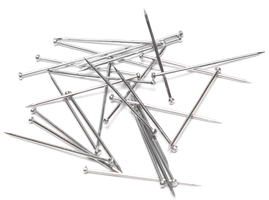
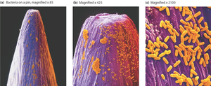
Rod-shaped bacterium: 0.5–1.0 μm wide, 1.0–4.0 μm long
Spherical (coccus) bacterium: 0.5–1.0 μm diameter
Microscopy in Microbiology
Introduction to the Microscope
The microscope is the most important tool for studying microorganisms. It allows visualization of structures too small for the naked eye.
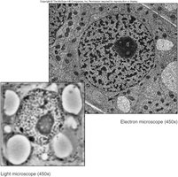
Light microscope: Uses visible light; magnifies up to ~1000x.
Electron microscope: Uses electron beams; magnifies >100,000x.
How a Microscope Works
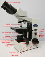
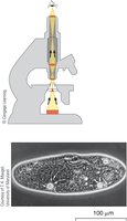
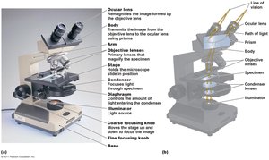
A compound microscope uses two sets of lenses (objective and ocular) to magnify specimens. The condenser lens focuses light onto the specimen but does not magnify.
Ocular lens: Remagnifies the image formed by the objective lens.
Objective lens: Primary magnification of the specimen.
Condenser lens: Focuses light onto the specimen; does not magnify.
Key Concepts in Microscopy
Magnification: Apparent increase in size of the specimen.
Resolution: Ability to distinguish two closely spaced objects as separate entities. High resolution is essential for clear images.
Contrast: Difference in light intensity between the specimen and background; improved by staining or adjusting light.
Limits of Resolution
The resolving power of a microscope determines the smallest distance between two points that can still be distinguished as separate. For bright-field microscopes, this is about 0.2 μm (200 nm).
Human eye: 0.1 mm
Light microscope: 0.2 μm
Electron microscope: 0.1 nm
Magnification without adequate resolution is meaningless.
Types of Microscopes
Different microscopes provide varying levels of detail. The bright-field, compound light microscope is the most commonly used in microbiology labs.
Microscope Setup and Use
Transport: Support the base with one hand and hold the arm with the other.
Light adjustment: Start with low intensity; increase as needed, especially for higher magnifications.
Eyepiece adjustment: Adjust distance for a single circular field of view.
Slide placement: Place specimen-side up and secure with slide holders.
Objective Lenses and Magnification
4x: Scanning objective (largest field of view)
10x: Low power
40x: High power, dry
100x: Oil immersion (must use immersion oil for clear focus)
Total magnification: Product of ocular lens (usually 10x) and objective lens (e.g., 10x × 40x = 400x)
Focusing and Parfocal Lenses
Begin focusing with 4x or 10x using the coarse adjustment knob.
Use fine focus for sharp images.
Microscopes are parfocal: once focused at low power, only minor adjustments are needed at higher powers.
Immersion Oil and Refraction
At 100x objective, immersion oil is used to prevent light refraction, which occurs when light passes from glass to air. Oil has a similar refractive index to glass, maintaining a straight light path and improving resolution.
Only the 100x objective lens should contact immersion oil.
Microscope Care and Handling
Clean the 100x objective lens before storing the microscope.
Lower the stage and return the objective lens to 4x before putting away.
Summary Table: Common Metric Units in Microbiology
Unit | Abbreviation | Value (in meters or liters) |
|---|---|---|
Centimeter | cm | 0.01 m |
Millimeter | mm | 0.001 m |
Micrometer | μm | 0.000001 m |
Nanometer | nm | 0.000000001 m |
Milliliter | ml | 0.001 l |
Microliter | μl | 0.000001 l |
Key Study Questions
Define genus and species; correctly write scientific names.
List and abbreviate common metric units.
State average sizes of bacillus- and coccus-shaped bacteria.
Explain magnification, resolution, and contrast in microscopy.
Describe microscope parts and their functions.
Calculate total magnification for different lens combinations.
Explain the use of immersion oil and the concept of parfocal lenses.
