 Back
BackMicrobiology Lab Review: Labs 1–6
Study Guide - Smart Notes
 Tailored notes based on your materials, expanded with key definitions, examples, and context.
Lab Safety and Aseptic Technique
Laboratory Safety Rules and Biosafety Levels (BSL)
Understanding and following laboratory safety rules is essential for preventing contamination and ensuring the safety of all personnel. Biosafety levels (BSL) classify laboratories based on the risk associated with the microorganisms handled:
BSL-1: Non-pathogenic microbes
BSL-2: Moderate-risk agents
BSL-3: Indigenous/exotic agents with potential for aerosol transmission
BSL-4: Dangerous/exotic agents with high risk of life-threatening disease (e.g., Ebola)
Example: Ebola virus is handled in BSL-4 laboratories due to its high pathogenicity and lack of treatment.
Key Safety Rule: Always wear appropriate personal protective equipment (PPE) and practice aseptic technique to prevent contamination.
Aseptic Technique
Aseptic technique refers to procedures used to prevent contamination of cultures, laboratory environment, and personnel. It is vital for accurate experimental results and safety.
Purpose: Prevent contamination of the microorganism, the environment, and personnel.
Application: Used in all microbiological procedures, including inoculation and media preparation.
Microscopy in Microbiology
Parts and Functions of the Microscope
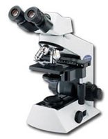
The compound light microscope is a fundamental tool in microbiology for observing microorganisms. Understanding its parts and their functions is essential for proper use.
Oculars (Eyepieces): Magnify the image, usually 10x.
Objectives: Provide additional magnification (e.g., 4x, 10x, 40x, 100x).
Stage: Holds the slide in place.
Coarse and Fine Focus: Adjust the focus of the image.
Condenser and Iris Diaphragm: Focus and regulate the amount of light.
Light Source: Illuminates the specimen.
Microscope Terminology
Total Magnification (TM): Calculated by multiplying the ocular lens magnification by the objective lens magnification.
Parfocal: The ability of the microscope to stay nearly in focus when switching between objectives.
Resolution: The ability to distinguish two points as separate entities.
Example Calculation: If the ocular lens is 4x and the objective is 20x, total magnification is .
Eukaryotic Microorganisms: Protozoa, Algae, and Fungi
Protozoan Characteristics
Protozoa are unicellular eukaryotes found in moist or aquatic environments. They lack cell walls and are enclosed by a plasma membrane or pellicle. Most are chemoheterotrophs, except Euglena, which is a photoautotroph. Protozoa reproduce mainly asexually and move by cilia, flagella, or pseudopodia.
Algae Characteristics
Algae are photosynthetic eukaryotes found in aquatic environments. They can be unicellular or multicellular, with cell walls typically composed of cellulose, though some have hard (silica) or soft (pectin) components. Most algae are photoautotrophs and reproduce sexually or asexually. Some are motile via flagella.
Fungal Characteristics
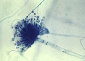
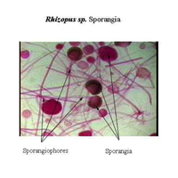
Fungi are found throughout the environment. Yeasts are unicellular, while molds are multicellular and composed of hyphae. Fungal cell walls contain chitin and may be septate or non-septate. Most fungi are saprotrophs, secreting enzymes to digest substrates externally before absorbing nutrients. Reproduction can be sexual or asexual.
Hypha: Long, thread-like structure forming the body of molds.
Chitin: Main component of fungal cell walls.
Microbial Culturing and Media
Five Basic Steps of Culturing Bacteria
Inoculation: Introduction of microbes into media.
Incubation: Providing optimal conditions for growth.
Isolation: Separating individual species.
Inspection: Observing colony morphology and characteristics.
Identification: Determining the species present.
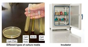
Types of Culture Media
Enriched Media: Contains nutrients to support fastidious organisms.
Differential Media: Distinguishes between organisms based on biochemical reactions.
Selective Media: Inhibits growth of some organisms while allowing others.
Reduced Media: Supports growth of anaerobes by reducing oxygen.
Inoculation Methods
Stabbing: Inoculating deep into agar to test for motility or oxygen requirements.
Stroke/Fishtail: Surface inoculation for growth observation.
Streak Plate Technique: Dilutes bacteria to isolate colonies.
Lawn Culture: Evenly spreading bacteria for antibiotic testing.
Quadrant Streaking: Isolates pure colonies from mixed cultures.
Colony Morphology (FACESS)
Form
Appearance
Color
Edges
Size
Surface
Mixed Culture: Contains two or more different microbial species.
Selective Media: Inhibits some bacteria while allowing others to grow.
Environmental Effects on Microbial Growth
Temperature Classification
Psychrophiles: -10 to 20°C (optimum 15°C)
Psychrotrophs: 0 to 30°C
Mesophiles: 15 to 45°C (optimum 37°C)
Thermophiles: 40 to 70°C (optimum 50°C)
Extreme Thermophiles: 65 to 110°C
High temperatures denature enzymes; low temperatures solidify membranes and inhibit enzyme function.
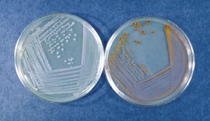
Chromogenesis
Chromogenesis is the production of pigment by microbes, which depends on enzyme activity and optimal temperature. For example, Serratia marcescens produces orange pigment at 25°C but not at 37°C.
pH and Microbial Growth
Acidophiles: pH 0–5.5
Neutrophiles: pH 5.5–8
Alkalinophiles: pH 7.5–14
pH affects enzyme activity and cell structure.
Osmotic Solutions
Hypertonic: Water leaves the cell, causing plasmolysis (cell shrivels).
Hypotonic: Water enters the cell, causing lysis (cell bursts).
Isotonic: No net water movement; cell remains unchanged.
Oxygen Requirements
Facultative Anaerobes: Can grow with or without oxygen.
Obligate Aerobes: Require oxygen.
Microaerophiles: Require low levels of oxygen.
Obligate Anaerobes: Cannot tolerate oxygen.
Aerotolerant: Do not use oxygen but tolerate its presence.
Staining Techniques in Microbiology
Types of Stains
Basic Stains: Contain a positive chromogen (e.g., methylene blue, crystal violet, safranin).
Acidic Stains: Contain a negative chromogen (e.g., nigrosin).
Staining Methods
Simple Stain: Uses one dye to reveal cell shape, size, and arrangement.
Differential Stain: Uses two dyes to distinguish cell types or structures (e.g., Gram stain, endospore stain, acid-fast stain).
Special Stains: Capsule, flagellar, and negative stains for specific structures.
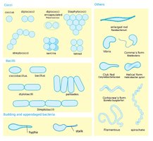
Simple Stain Example
Simple stains such as methylene blue allow visualization of bacterial morphology under the microscope.
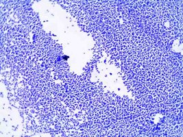
Negative Stain
Negative staining uses an acidic dye (e.g., nigrosin) to stain the background, not the cells. It is useful for observing live bacteria that cannot withstand heat fixing and for determining size, shape, and arrangement.
Bacterial Shapes and Arrangements
Cocci: Spherical
Bacilli: Rod-shaped
Spiral: Spiral-shaped
Arrangements include single, pairs (diplo-), chains (strepto-), clusters (staphylo-), etc.
Glossary of Key Terms
Ubiquity: Presence of microbes everywhere.
Chromogenesis: Pigment production by microbes.
Colony Morphology: Observable characteristics of microbial colonies.
Pure Culture: Culture containing only one species of microorganism.
