 Back
BackMicrobiology Lab Techniques: Staining, Biochemical Tests, and Selective Media
Study Guide - Smart Notes
 Tailored notes based on your materials, expanded with key definitions, examples, and context.
Staining Techniques in Microbiology
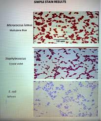
Simple Staining
Simple stains are used to visualize bacterial cells and determine their morphology, arrangement, and size. These stains use a single basic dye that binds to negatively charged bacterial cell components, making cells visible against a light background.
Methylene Blue: Stains bacterial cells blue. Useful for observing general cell shape and arrangement.
Crystal Violet: Stains cells purple. Also serves as the primary stain in Gram staining.
Safranin: Stains cells pink/red. Used as a counterstain in Gram and endospore staining.
Application: Simple stains help distinguish between cocci, bacilli, and other bacterial shapes.
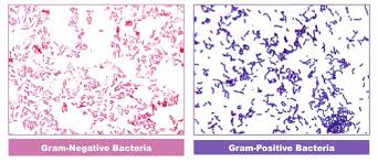
Gram Staining
Gram staining is a differential staining technique that classifies bacteria into Gram-positive and Gram-negative groups based on cell wall structure. The process involves four main steps:
Crystal Violet (Primary Stain): Stains all cells purple.
Iodine (Mordant): Forms a complex with crystal violet, trapping it in the cell wall.
Alcohol/Acetone (Decolorizer): Removes stain from Gram-negative cells, which have thinner peptidoglycan layers.
Safranin (Counterstain): Stains decolorized Gram-negative cells pink/red.
Gram-positive bacteria: Retain crystal violet and appear purple.
Gram-negative bacteria: Lose crystal violet and take up safranin, appearing pink/red.
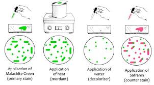
Endospore Staining
Endospore staining is a differential technique used to detect bacterial endospores, which are resistant structures formed by some genera (e.g., Bacillus, Clostridium). The process uses malachite green and heat to stain spores, followed by safranin to stain vegetative cells.
Malachite Green + Heat: Stains endospores green.
Water Rinse: Removes stain from vegetative cells.
Safranin: Counterstains vegetative cells pink/red.
Endospores: Green
Vegetative cells: Pink/red
Biochemical Tests for Bacterial Identification
Catalase Test
The catalase test detects the presence of the enzyme catalase, which breaks down hydrogen peroxide into water and oxygen. Bubbles indicate a positive result.
Positive: Immediate bubbling (e.g., Staphylococcus species)
Negative: No bubbles (e.g., Streptococcus species)
Reaction:
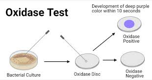
Oxidase Test
The oxidase test identifies bacteria that produce cytochrome c oxidase, an enzyme in the electron transport chain. A dark purple color within 10–30 seconds indicates a positive result.
Positive: Dark purple color (e.g., Pseudomonas)
Negative: No color change (e.g., Enterobacteriaceae)
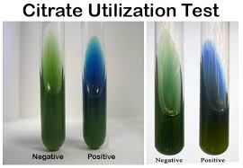
Citrate Utilization Test
This test determines if an organism can use citrate as its sole carbon source. Utilization of citrate raises the pH, turning the bromothymol blue indicator from green to blue.
Positive: Blue color with growth
Negative: Green color, no growth
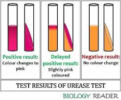
Urea Hydrolysis Test
The urea hydrolysis test detects the enzyme urease, which breaks down urea into ammonia and carbon dioxide. Ammonia raises the pH, causing the phenol red indicator to turn bright pink.
Positive: Bright pink color
Negative: Yellow/orange color
Reaction:
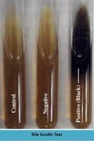
Bile Esculin Test
The bile esculin test identifies bacteria that can hydrolyze esculin in the presence of bile. A positive result is indicated by blackening of the medium.
Positive: Black medium (e.g., Enterococcus species)
Negative: No blackening
Selective and Differential Media
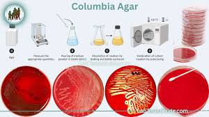
Columbia CNA Agar
Columbia CNA agar contains colistin and nalidixic acid to inhibit Gram-negative bacteria, allowing the growth of Gram-positive organisms. The addition of 5% sheep blood enables observation of hemolysis patterns.
Selects for: Gram-positive bacteria
Hemolysis patterns: Beta (clear zone), Alpha (greenish zone), Gamma (no hemolysis)
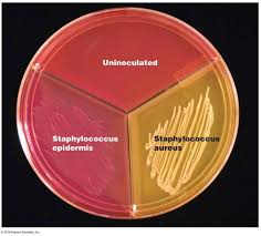
Mannitol Salt Agar (MSA)
MSA is both selective and differential. Its high salt concentration selects for salt-tolerant bacteria (mainly Staphylococcus species). Mannitol fermentation is indicated by a yellow color change in the medium due to acid production.
Positive (yellow): Staphylococcus aureus (mannitol fermenter)
Negative (pink/red): Staphylococcus epidermidis (non-fermenter)
MacConkey Agar
MacConkey agar is selective for Gram-negative enteric bacteria and differentiates lactose fermenters from non-fermenters. Bile salts and crystal violet inhibit Gram-positive bacteria. Lactose fermenters produce pink/red colonies due to acid production.
Positive (pink/red colonies): Lactose fermenters (e.g., Escherichia coli)
Negative (colorless colonies): Non-lactose fermenters (e.g., Salmonella)

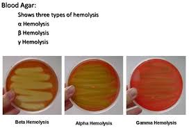
Blood Agar and Hemolysis Patterns
Blood agar is a differential medium used to distinguish bacteria based on their hemolytic properties:
Beta hemolysis: Complete lysis of red blood cells (clear zone)
Alpha hemolysis: Partial lysis (greenish zone)
Gamma hemolysis: No lysis (no change)
Antibiotic Sensitivity Testing
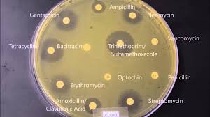
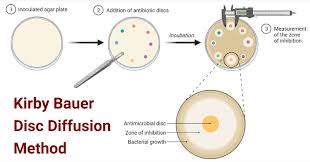
Kirby-Bauer Disk Diffusion Test
The Kirby-Bauer test evaluates bacterial sensitivity to antibiotics. Disks containing antibiotics are placed on an agar plate inoculated with bacteria. After incubation, zones of inhibition (clear areas) are measured to determine susceptibility.
Large zone: Sensitive to antibiotic
Small/no zone: Resistant to antibiotic
Bacterial Transformation (pGLO)
Genetic Transformation Using Plasmid DNA
Bacterial transformation involves introducing foreign DNA (plasmid) into bacteria, conferring new traits such as antibiotic resistance or fluorescence. The pGLO system uses a plasmid with a gene for green fluorescent protein (GFP) and an antibiotic resistance marker.
Add plasmid DNA to competent bacterial cells.
Heat shock to facilitate DNA uptake.
Plate cells on selective media (e.g., containing ampicillin).
Observe for growth and fluorescence under UV light.
Growth on antibiotic plate: Indicates successful transformation.
Green fluorescence: Indicates expression of GFP gene.
Summary Table: Key Tests and Media
Test / Media | Positive Result | Selects For / Meaning |
|---|---|---|
Gram Stain | Gram+ purple, Gram− pink | Differentiates by cell wall |
Endospore Stain | Green spores, red cells | Detects endospores |
Catalase | Bubbles | Detects catalase enzyme |
Oxidase | Purple | Detects cytochrome c oxidase |
Citrate | Blue | Organism uses citrate |
Urea | Bright pink | Detects urease enzyme |
Phenylalanine | Green | Detects phenylalanine deaminase |
Decarboxylase | Purple | Detects amino acid decarboxylation |
Bile Esculin | Black | Esculin hydrolysis |
ELISA | Color change | Antigen/antibody detection |
Kirby-Bauer | Large clear zone | Antibiotic sensitivity |
Quick Memory Tricks
Blue = citrate used
Pink pee = urease positive
Mac pink = lactose fermenter
MSA yellow = mannitol fermented
Black bile esculin = positive
Catalase = bubbles
Oxidase = purple
Green spores survive
