 Back
BackMicrobiology Laboratory Study Guide: Techniques, Morphology, and Biochemical Assays
Study Guide - Smart Notes
 Tailored notes based on your materials, expanded with key definitions, examples, and context.
Laboratory Practica and Station Format
Lab Practica Structure
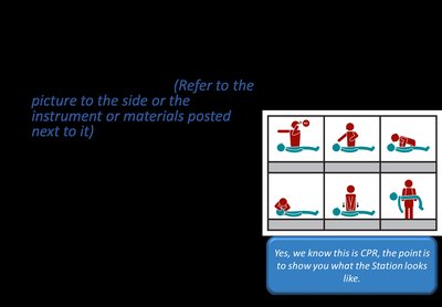
Microbiology laboratory practica are hands-on, station-based exams designed to assess practical skills and knowledge. Each station focuses on a specific lab technique, protocol, or concept, and students rotate through stations under timed conditions.
Station Arrangement: The lab is divided into two halves, each with 16 stations. Students start at different stations and rotate sequentially.
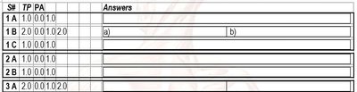
Question Format: Each station presents 2-4 questions related to objectives, methods, results, and importance of the lab technique.
Answer Sheets: Students record answers in designated spaces, noting station numbers and points earned.


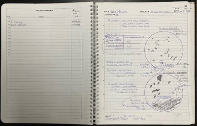
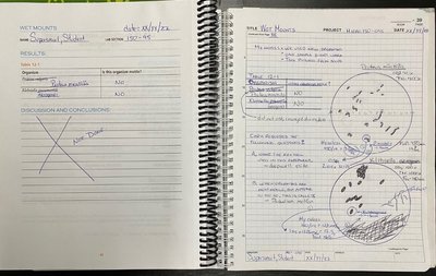
Laboratory Notebook Guidelines
Notebook Organization and Data Collection
A laboratory notebook (LN) is essential for recording all experimental data, observations, and notes. Proper organization and thorough documentation are critical for scientific accuracy and reproducibility.
Recording Data: All experimental data, measurements, drawings, and notes must be entered in pen (blue or black).
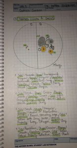
Drawings: Microscope drawings should include measurements and scale bars.
Organization: Maintain a clear table of contents and label all entries.
Corrections: Make corrections clearly and transfer fidelity between original and carbon copy pages.
Laboratory Safety and Clean-Up
Safety Protocols
Strict adherence to laboratory safety protocols is required to prevent accidents and contamination. Personal protective equipment (PPE) and proper disposal of materials are mandatory.
PPE: Wear lab coats, gloves, closed-toe shoes, and tie back hair.
Disposal: Dispose of glassware, gloves, and biohazard materials in designated containers.
Clean-Up: Clean benches, wash glassware, and remove all labels before leaving the lab.

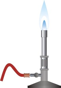
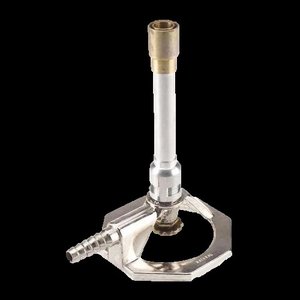
Bunsen Burner Anatomy and Use
Bunsen Burner Components and Flame Types
The Bunsen burner is a fundamental tool for sterilization and heating in microbiology labs. Understanding its anatomy and flame characteristics is essential for safe and effective use.
Components: Base, barrel, collar, gas tap, oxygen intake, and gas valve regulator.
Flame Types: Orange/yellow (cool, luminous), blue (hot, non-luminous). The tip of the inner blue cone is the hottest and used for sterilization.
Safety: Never leave unattended; keep away from flammable materials.

Bacterial Morphology
Micro-Morphology: Shapes and Arrangements
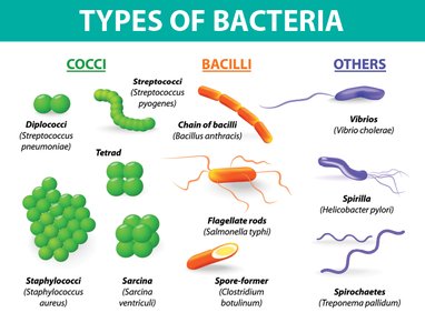
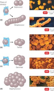
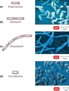
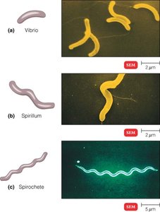
Bacterial cells exhibit distinct shapes and arrangements, which are key to identification and classification.
Coccus: Spherical; arrangements include diplococci, streptococci, tetrads, sarcinae, and staphylococci.
Bacillus: Rod-shaped; arrangements include single bacilli, diplobacilli, streptobacilli, and coccobacilli.
Spiral: Includes vibrios (curved rods), spirilla (rigid helices), and spirochetes (flexible helices).
Macro-Morphology: Colony Characteristics
Colony Morphology on Plates
Colony morphology refers to the observable characteristics of microbial colonies grown on solid media. These features aid in identification and differentiation.
Form: Punctiform, circular, filamentous, rhizoid, irregular, spindle.
Elevation: Flat, raised, convex, pulvinate, umbonate, crateriform.
Margin: Entire, undulate, filamentous, lobate, erose, curled, scalloped.
Surface: Shiny, dull, smooth, veined, rough.
Texture: Moist, brittle, butyrous, viscid.
Color: Opaque, cloudy, translucent, iridescent.
Aseptic Techniques and Isolation of Bacteria
Aseptic Technique Principles
Aseptic technique is essential for minimizing contamination and maintaining pure cultures. It involves careful handling of samples, instruments, and media.
Objectives: Minimize contamination, maintain pure cultures, ensure valid results.
Steps: Sterilize instruments (incineration), proper tube manipulation, flame tubes before and after use, careful transfer from broth, slant, or plate.
Inoculation: Use correct techniques for broth, slant, and plate inoculation (swirl, fishtail, streak).
Biochemical Activity of Microorganisms
Metabolic Assays and Media
Biochemical assays are used to identify microorganisms based on their metabolic activities. These tests utilize specialized media and reagents to detect enzymatic reactions and fermentation.
Common Assays: Casein hydrolysis, starch hydrolysis, carbohydrate fermentation, amino acid decarboxylation, DNase, hemolysin, coagulase, phenylalanine deaminase, H2S production, IMViC tests, catalase, oxidase.
Media Types: TSA, SMA, EMB, MSA, fermentation broths, decarboxylation broths, DNase agar, blood agar, phenylalanine agar, TSI slants.
Interpretation: Observe color changes, zones of clearing, gas production, and other indicators to determine positive or negative results.
Staining Techniques
Principles and Protocols
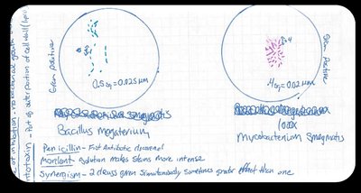
Staining is used to visualize and differentiate microorganisms under the microscope. Common stains include simple stains, negative stains, Gram stain, endospore stain, and acid-fast stain.
Simple Stain: Uses a single dye to color cells for basic observation.
Negative Stain: Stains the background, leaving cells unstained; useful for capsules.
Gram Stain: Differentiates bacteria into Gram-positive (purple) and Gram-negative (pink/red) based on cell wall structure.
Endospore Stain: Identifies spore-forming bacteria using malachite green and safranin.
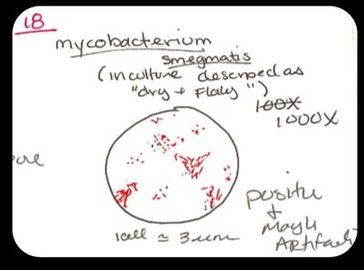
Acid-Fast Stain: Detects mycolic acid-containing bacteria (e.g., Mycobacterium spp.) using carbol-fuchsin and methylene blue.
Microscopy and Measurement
Microscope Use and Field of View Calculations
Proper use of the microscope is essential for observing microorganisms. Measurements are made using the field of view (FoV) and scale bars.
Objectives: 4x, 10x, 40x, 100x (oil immersion).
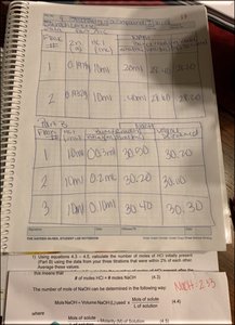
FoV Calculation: Use the formula , where SS is specimen size, Target is number of squares occupied, FoV size is diameter, and D is total squares across diameter.
Drawing: Always include measurements and scale bars in microscope drawings.
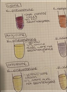
Biochemical Assay Table: Decarboxylation Test Results
Substrate | Organism | Result | Interpretation |
|---|---|---|---|
Lysine | K. pneumoniae | Dark purple/violet | Positive for decarboxylation |
Arginine | K. pneumoniae | Yellow | Negative; amino acids not decarboxylated |
Ornithine | K. pneumoniae | Yellow/some brown | Negative; amino acids not decarboxylated |
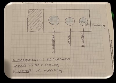
Example: Catalase Test Results
Organism | Result | Interpretation |
|---|---|---|
S. pyogenes | No bubbling | Catalase negative |
M. luteus | No bubbling | Catalase negative |
B. cereus | Bubbling | Catalase positive |
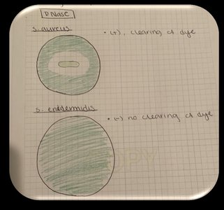
Example: DNase Test Results
Organism | Result | Interpretation |
|---|---|---|
S. aureus | Clearing of dye | DNase positive |
S. epidermidis | No clearing of dye | DNase negative |
Summary
This guide covers essential microbiology laboratory techniques, including safety, aseptic methods, bacterial morphology, staining, microscopy, and biochemical assays. Proper documentation, observation, and interpretation of results are critical for success in laboratory practica and exams.
