 Back
BackMicrobiology Laboratory Techniques and Concepts: Lab Exam 1 Study Guide
Study Guide - Smart Notes
 Tailored notes based on your materials, expanded with key definitions, examples, and context.
Microbiological Media and Culture Techniques
Preparation and Types of Microbiological Media
Microbiological media are essential for the cultivation and study of microorganisms. Media can be solid or liquid, depending on the presence of a solidifying agent such as agar.
Solid Media: Contains agar, used for isolating and culturing bacteria.
Liquid Media: Lacks agar, used for growing bacteria in suspension.
Sterilization: Media must be sterilized before use, typically by autoclaving at 121°C, 15 psi for 15 minutes.
Example: Sabouraud Dextrose Agar is a solid medium with a final pH of 5.6 and 4% dextrose concentration.
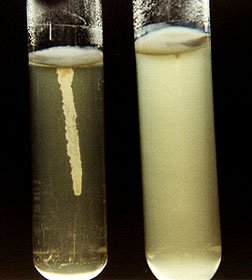
Inoculation and Aseptic Techniques
Aseptic technique is critical for preventing contamination during the transfer and inoculation of cultures. Different tools and methods are used depending on the type of culture.
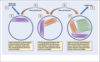
Streak Plate Method: Used to isolate single colonies from a mixed culture.
Agar Slant: Used for maintaining pure cultures over extended periods.
Broth Culture: Used for growing bacteria in liquid form.
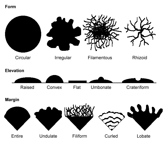
Describing Growth: Terms such as turbidity, pellicle, and sediment are used for broth cultures; form, elevation, and margin describe colonies on solid media.
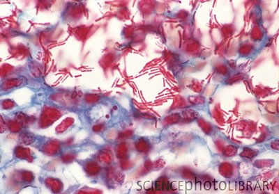
Bacterial Staining Procedures
Gram Stain
The Gram stain is a differential staining technique used to classify bacteria based on cell wall structure.
Reagents (in order): Crystal Violet (primary stain), Iodine (mordant), Ethanol (decolorizer), Safranin (counterstain).
Gram Positive: Thick peptidoglycan layer retains crystal violet-iodine complex (purple).
Gram Negative: Thin peptidoglycan and lipid membrane lose primary stain after alcohol (red after counterstain).
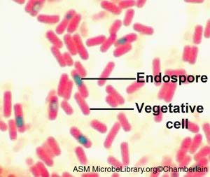
Schaffer-Fulton Endospore Stain
Reagents: Malachite Green (primary stain), Safranin (counterstain).
Endospores: Stain green; vegetative cells stain red.
Acid-Fast Stain (Kinyoun Method)
Reagents: Carbol Fuchsin (primary stain), Acidified alcohol (decolorizer), Brilliant Green or Methylene Blue (counterstain).
Acid-Fast Cells: Appear red due to mycolic acid in cell wall.
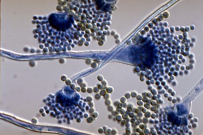
Medical Mycology
Histoplasma capsulatum
Histoplasma capsulatum produces tuberculated chlamydospores for reproduction and dispersal. The disease caused is histoplasmosis, acquired by inhalation of spores, commonly found in the Ohio and Mississippi River Valleys.
Aspergillus and Aflatoxin
Aspergillus is an opportunistic pathogen producing aflatoxin, which can cause liver necrosis and cancer. It primarily causes respiratory infections (aspergillosis).
Transmission of Microorganisms
Hand Washing and Disease Prevention
Hand washing removes transient microbes but does not eliminate all bacteria. Surgical scrubbing reduces contamination but does not sterilize skin. Blood agar is an enriched and differential medium used to culture fastidious organisms and observe hemolytic patterns. Transmission via fomites (e.g., drinking glass rim) is a common route for infection.
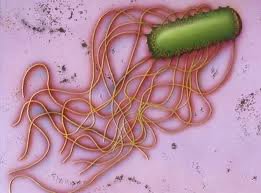
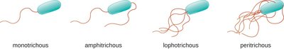
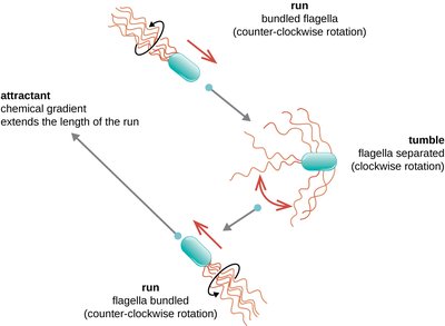
Bacterial Motility
Determining Motility
Bacterial motility can be observed using semisolid motility medium, flagella stains, or hanging drop slides. Most bacteria are motile via flagella, while spirochetes use axial filaments.
Gram Positive Spore-forming Bacteria
Bacillus and Clostridium
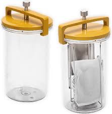
Gram positive spore-forming bacteria include Bacillus (aerobic) and Clostridium (anaerobic). Diseases caused include anthrax, botulism, tetanus, gas gangrene, and antibiotic-resistant diarrhea. Anaerobic conditions are provided by Gas Pack jars, cooked meat medium, thioglycollate medium, and anaerobic incubators. Candle jars are used for capnophiles, not strict anaerobes.
Destruction and Control of Microorganisms
Physical and Chemical Methods
Microbial control methods include refrigeration (bacteriostatic), autoclaving (bacteriocidal), dry oven, and boiling. Endospores are resistant to heat and require autoclaving for destruction.
Procedure | Temperature (°C) | Time (min) | Pressure (psi) |
|---|---|---|---|
Autoclave | 121 | 15 | 15 |
Oven | 160-170 | 120-180 | -- |
Boiling | 100 | 15-20 | -- |
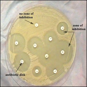
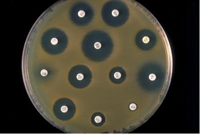
Antibiotic Susceptibility Testing
The Kirby Bauer Disk Susceptibility Test is used to determine bacterial sensitivity to antibiotics. Zones of inhibition are measured and compared to standard tables to interpret results as sensitive, intermediate, or resistant. The size of the zone depends on the diffusion rate of the antibiotic, not necessarily its effectiveness.
Mycobacterium and Acid-Fast Staining
Kinyoun Acid Fast Stain Procedure
Mycobacteria are distinguished by their acid-fast cell walls containing mycolic acid. The Kinyoun method stains acid-fast cells red, while non-acid-fast cells are counterstained green or blue. Diseases include tuberculosis (Mycobacterium tuberculosis) and leprosy (Mycobacterium leprae).
Summary Table: Colony Morphology Terms
Term | Description | Examples |
|---|---|---|
Form | Overall shape of colony | Circular, Irregular, Filamentous, Rhizoid |
Elevation | Side view appearance | Raised, Convex, Flat, Umbonate, Crateriform |
Margin | Edge detail | Entire, Undulate, Filiform, Curled, Lobate |
Additional info: Academic context and explanations have been expanded for clarity and completeness. All images included are directly relevant to the adjacent content and reinforce key laboratory concepts.
