 Back
BackMicrobiology Practical Review: Essential Laboratory Techniques and Concepts
Study Guide - Smart Notes
 Tailored notes based on your materials, expanded with key definitions, examples, and context.
Microbiology Laboratory Safety and Clean-Up
Lab Safety Guidelines
Proper laboratory safety is fundamental in microbiology to prevent contamination and ensure the safety of all personnel. These guidelines must be followed at all times:
Lab coat: Wear at all times in the lab.
Hand hygiene: Wash hands before and after lab work.
Disinfection: Disinfect lab benches before and after lab.
Personal protection: Cover all cuts and scrapes; tie back long hair.
Microbe transfer: Avoid inhaling airborne microbes during transfers.
Culture handling: Never lay culture tubes on the bench.
Accident reporting: Report all spills and accidents immediately.
Disposal: Infectious materials and gloves go in biohazard trash.
Clean-Up Procedures
Petri dishes and small contaminated items: Dispose in biohazard trash.
Broken glass: Use designated container (no paper towels).
Slides: Clean oil off prepared slides with Kim wipes; plain glass slides with Bon Ami (wear goggles).
Test tubes: Remove markings/tape, loosen caps, boil for 15 minutes, pour contents into biohazard trash, wash tubes thoroughly.
Decontamination: Cover spills with disinfectant, inform neighbors and instructor, wait 15 minutes, then clean up.
Microbiological Equipment and Techniques
Bunsen Burner Usage
The Bunsen burner is essential for sterilization and aseptic technique. Oxygen flow is controlled by rotating the barrel:
Clockwise: Less oxygen
Counterclockwise: More oxygen
Key parts: barrel, collar, base, gas valve intake.
Preparing Agar Plates
Agar plates are used to culture microbes. Agar talls (solid media in test tubes) are melted, cooled, and poured into petri dishes. The agar solidifies in about 15 minutes.
Steam Slide Preparation
Steam is used for endospore and acid-fast staining. Slides are heat-fixed, placed on a steaming coffee can, covered with a paper towel, and stained for 10 minutes.
Microbiological Media and Their Uses
Types of Media
Tryptic Soy Agar (TSA): General purpose, neutral pH, supports a wide range of microbes.
Blood Agar Plate (BAP): TSA with sheep's blood, used to detect hemolysis.
Rodac Plates: Raised agar for surface sampling.
Hemolysis Types
Alpha hemolytic: Partial hemolysis, brownish discoloration.
Beta hemolytic: Complete hemolysis, clearing of red color.
Gamma hemolytic: No hemolysis, no change in media.
Bacterial Morphology and Colony Characteristics
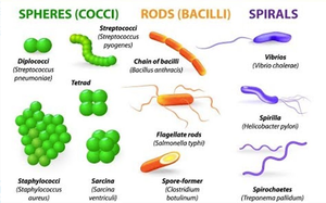
Bacterial Cell Morphologies
Bacteria exhibit three basic shapes:
Cocci: Spherical
Bacilli: Rod-shaped
Spirals: Curved or spiral-shaped (vibrios, spirilla, spirochetes)
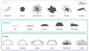
Colony Morphology
Colony morphology is used to identify bacteria based on appearance:
Shape: Circular, irregular, filamentous, etc.
Margin: Entire, undulate, lobate, curled, etc.
Elevation: Flat, raised, convex, umbonate, etc.
Aseptic Technique and Pure Culture Isolation
Aseptic Methods
Aseptic technique prevents contamination and exposure. Pure cultures contain only one organism, while mixed cultures contain multiple.
Flame sterilization: Use the hottest part of the flame (tip of blue inner cone).
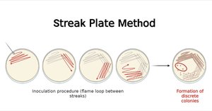
Streak Plate Method
The streak plate method is used to isolate pure cultures from mixed samples. It involves streaking bacteria across the plate in a specific pattern to separate individual colonies.
Microscopy in Microbiology
Bright-Field Microscope Structure
Microscopes are essential for observing microorganisms. Key parts include:
Ocular/eyepiece: 10x magnification
Objective lenses: 4x, 10x, 40x, 100x
Stage, arm, mechanical stage adjustment, condenser
Total magnification:
Microscopes are parfocal, meaning focus remains consistent when switching objectives.
Omnipresence of Microorganisms
Microbes in the Environment
Bacteria are found everywhere. Rodac plates are used to sample surfaces, while TSA plates capture airborne microbes.
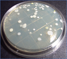
Human Microbiome and Bacterial Pigments
Human Microbiome
Humans host a variety of microorganisms, many of which are beneficial. TSA, TSB, and blood agar plates are used to culture skin and throat microbes, and to test for hemolytic bacteria.
Bacterial Pigments
Some bacteria produce pigments, aiding identification. Gram-positive bacteria often produce pigments regardless of conditions, while Gram-negative bacteria like Serratia marcescens depend on incubation conditions.
Bacterial Motility
Motility Studies
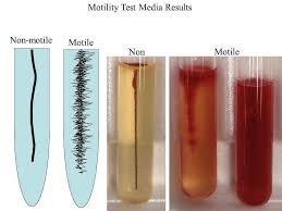
Motile bacteria can move, while Brownian motion is random movement caused by water molecules. Wet mounts help distinguish true motility from Brownian motion.
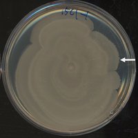
Motility Media and Swarming
Motility media deeps are stabbed with inoculation needles to test for motility. Highly motile bacteria exhibit swarming on solid media, forming concentric circles.
Staining Techniques in Microbiology
Simple Staining
Staining increases contrast for microscopic observation. Simple stains use one dye; direct stains color cells, indirect stains color the background.
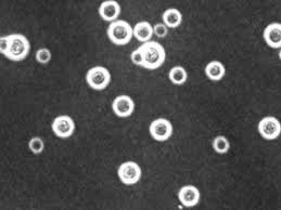
Negative Staining and Capsule Detection
Negative staining (using India ink) highlights bacterial capsules. Counterstains (safranin or crystal violet) color the cell, leaving the capsule clear.
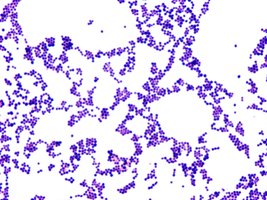
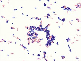
Gram Staining
Gram staining differentiates bacteria by cell wall type:
Gram positive: Dark blue/purple (retains crystal violet)
Gram negative: Light red/pink (retains safranin)
Steps: Primary stain (crystal violet), mordant (iodine), decolorizer (alcohol), counterstain (safranin).
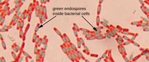
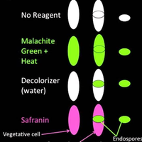
Endospore Staining
Endospores are highly resistant structures. The Schaeffer-Fulton method uses steam and malachite green to stain endospores, with safranin as a counterstain.
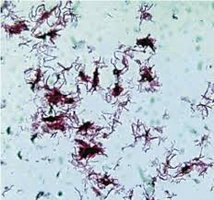
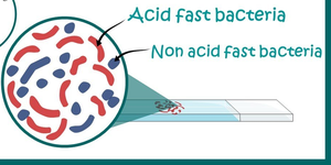
Acid-Fast Staining
Acid-fast staining (Ziehl–Neelsen method) identifies bacteria with mycolic acid in their cell walls (e.g., Mycobacteria). Acid-fast cells appear red; non-acid-fast cells appear blue.
Summary Table: Colony Morphology
The following table summarizes key colony morphology characteristics:
Shape | Margin | Elevation |
|---|---|---|
Circular, Irregular, Filamentous | Entire, Undulate, Lobate, Curled | Flat, Raised, Convex, Umbonate |
Additional info: Colony morphologies are used for preliminary identification of bacterial species. | Additional info: Margins help distinguish between species. | Additional info: Elevation is often noted in lab reports. |
Key Equations
Total Magnification:
