 Back
BackMicrobiology Practical Review: Media, Indicators, and Antimicrobial Susceptibility
Study Guide - Smart Notes
 Tailored notes based on your materials, expanded with key definitions, examples, and context.
Q1. What is the difference between a pH indicator and an indicator molecule? How are these applied in microbiology media?
Background
Topic: Indicators in Microbiology Media
This question tests your understanding of how different types of indicators are used in microbiological media to detect metabolic changes or the presence of specific products.
Key Terms and Concepts:
pH Indicator: A chemical that changes color depending on the pH of the environment (e.g., phenol red, methyl red).
Indicator Molecule: Any molecule that signals a biochemical reaction, not limited to pH changes (e.g., Kovac’s reagent for indole detection).
Step-by-Step Guidance
Review the definition of a pH indicator and how it is used to detect acid or base production in media (e.g., phenol red in carbohydrate fermentation broths).
Compare this to indicator molecules that may detect other metabolic products (e.g., indole, H2S, or starch hydrolysis).
Think about examples from your lab manual where each type is used and what color changes or reactions you would expect.
Try solving on your own before revealing the answer!
Q2. How do you determine if a medium is selective, differential, or both? Apply this to MSA, SIM, Starch Hydrolysis, Phenol Red broth, EMB, MacConkey’s, MRVP, and Urease media.
Background
Topic: Types of Microbiological Media
This question tests your ability to classify media based on their function and to apply this knowledge to specific examples.
Key Terms:
Selective Media: Inhibits growth of some organisms while allowing others to grow.
Differential Media: Distinguishes between organisms based on biochemical reactions, often with color changes.
Both: Some media can be both selective and differential (e.g., MSA, EMB, MacConkey’s).
Step-by-Step Guidance
For each medium, identify the selective agent (if any) and the differential indicator.
Determine which organisms are selected for or against, and what reactions are differentiated (e.g., fermentation, enzyme production).
Use your lab manual to match each medium to its category and note the expected results for common organisms.
Try solving on your own before revealing the answer!
Q3. What type of bacteria does Mannitol Salt Agar (MSA) help us isolate? Is it selective, differential, or both? How do you know?
Background
Topic: Mannitol Salt Agar (MSA)
This question focuses on understanding the purpose and function of MSA in isolating and differentiating bacteria, especially Staphylococcus species.
Key Terms and Concepts:
Selective Agent: High salt concentration (7.5% NaCl)
Differential Agent: Mannitol and phenol red (pH indicator)
Step-by-Step Guidance
Recall which bacteria can tolerate high salt concentrations (e.g., Staphylococcus spp.).
Consider how mannitol fermentation is detected (color change in phenol red from red to yellow).
Decide if MSA is selective, differential, or both, and explain your reasoning based on its components and expected results.
Try solving on your own before revealing the answer!
Q4. What is the indicator molecule used in detecting indole? What does the indole portion of the SIM tube test for?
Background
Topic: Indole Test in SIM Medium
This question tests your knowledge of the biochemistry behind the indole test and the reagents used.
Key Terms and Concepts:
SIM Medium: Tests for Sulfide, Indole, and Motility.
Indole Test: Detects the breakdown of tryptophan to indole.
Indicator Molecule: Kovac’s reagent.
Step-by-Step Guidance
Recall the metabolic pathway: tryptophanase breaks down tryptophan to indole.
Identify the reagent added after incubation (Kovac’s reagent) and the color change that indicates a positive result.
Explain what a positive indole test means about the organism’s metabolism.
Try solving on your own before revealing the answer!
Q5. What tests form a black precipitate, and what does this indicate?
Background
Topic: H2S Production in Microbiology
This question focuses on identifying tests that detect hydrogen sulfide production and interpreting the results.
Key Terms and Concepts:
Black Precipitate: Indicates H2S production (e.g., SIM, TSI agar).
Mechanism: H2S reacts with iron salts to form black iron sulfide.
Step-by-Step Guidance
List the media that test for H2S production (e.g., SIM, TSI).
Describe the chemical reaction that leads to black precipitate formation.
Interpret what a positive result means about the organism’s metabolism.
Try solving on your own before revealing the answer!
Q6. What is the enzyme that allows for the breakdown of starch? What would that do to a starch-infused media? How would it look if we were to add iodine?
Background
Topic: Starch Hydrolysis Test
This question tests your understanding of exoenzyme activity and the interpretation of starch hydrolysis results.
Key Terms and Concepts:
Enzyme: Amylase
Indicator: Iodine (forms blue-black complex with starch)
Step-by-Step Guidance
Recall the function of amylase in breaking down starch.
Describe what happens when iodine is added to the plate (color change where starch is present).
Explain how to interpret a positive versus negative result.
Try solving on your own before revealing the answer!
Q7. What does the MR portion of the MRVP test for? Why is methyl red used instead of phenol red? What are phenol red and methyl red considered? What about the VP portion?
Background
Topic: MRVP Test
This question tests your understanding of the metabolic pathways detected by the MRVP test and the role of pH indicators.
Key Terms and Concepts:
MR (Methyl Red) Test: Detects mixed acid fermentation.
VP (Voges-Proskauer) Test: Detects acetoin production.
Indicators: Methyl red (for MR), Barritt’s reagents (for VP).
Step-by-Step Guidance
Explain why methyl red is used (detects lower pH than phenol red).
Describe what a positive MR or VP result indicates about the organism’s metabolism.
Classify methyl red and phenol red as pH indicators.
Try solving on your own before revealing the answer!
Q8. What is the name of the inverted tube inside the phenol red broth tubes?
Background
Topic: Gas Production in Fermentation Tests
This question tests your knowledge of how gas production is detected in fermentation broths.
Key Terms and Concepts:
Durham Tube: Small inverted tube used to capture gas produced during fermentation.
Step-by-Step Guidance
Recall the purpose of the Durham tube in detecting gas production.
Describe how you would interpret the presence of a bubble in the tube.
Try solving on your own before revealing the answer!
Q9. How do you interpret results from a table of organisms and media reactions (e.g., MSA, SIM, SC, H2S)?
Background
Topic: Interpreting Microbiology Results Tables
This question tests your ability to analyze and draw conclusions from experimental data presented in tables.
Key Terms and Concepts:
Growth (+/-): Indicates whether the organism can grow on the medium.
Color Change (+/-): Indicates a positive or negative biochemical reaction.
Step-by-Step Guidance
For each organism, note the Gram reaction and which media it can grow on.
Interpret what a positive or negative result means for each test (e.g., color change, growth, motility).
Apply this reasoning to predict outcomes for unknown organisms or new combinations.
Try solving on your own before revealing the answer!
Q10. What are the three types of hemolysis observed on blood agar? What kind of organisms are usually grown on blood agar?
Background
Topic: Hemolysis on Blood Agar
This question tests your knowledge of hemolytic patterns and their significance in identifying bacteria.
Key Terms and Concepts:
Alpha Hemolysis: Partial hemolysis, greenish color (e.g., Streptococcus pneumoniae).
Beta Hemolysis: Complete hemolysis, clear zone (e.g., Streptococcus pyogenes).
Gamma Hemolysis: No hemolysis.
Blood Agar: Used to grow fastidious organisms, especially Streptococcus species.
Step-by-Step Guidance
List the three types of hemolysis and describe their appearance on blood agar.
Identify which organisms are commonly tested using blood agar.
Try solving on your own before revealing the answer!
Q11. What are coliforms?
Background
Topic: Coliform Bacteria
This question tests your understanding of the definition and significance of coliforms in microbiology.
Key Terms and Concepts:
Coliforms: Gram-negative, lactose-fermenting rods associated with fecal contamination.
Step-by-Step Guidance
Recall the characteristics that define coliforms.
Explain why coliforms are important indicators in water and food safety testing.
Try solving on your own before revealing the answer!
Q12. How do non-ionizing and ionizing radiation work in microbial control? Are UV lamps used in lab non-ionizing? Can UV rays bypass the lid?
Background
Topic: Radiation in Microbial Control
This question tests your understanding of physical methods for controlling microbial growth.
Key Terms and Concepts:
Non-ionizing Radiation: UV light, causes DNA damage (thymine dimers).
Ionizing Radiation: X-rays, gamma rays, cause more severe DNA damage.
UV Lamps: Non-ionizing, limited penetration (cannot pass through lids or plastic).
Step-by-Step Guidance
Describe the mechanism of action for non-ionizing and ionizing radiation.
Explain the limitations of UV light in laboratory disinfection.
Try solving on your own before revealing the answer!
Q13. How do you interpret antibiotic disk diffusion results? What does sensitive versus resistant look like on a plate?
Background
Topic: Antibiotic Susceptibility Testing (Kirby-Bauer Disk Diffusion)
This question tests your ability to interpret the results of antibiotic susceptibility testing using disk diffusion.
Key Terms and Concepts:
Zone of Inhibition: Clear area around an antibiotic disk where bacteria do not grow.
Sensitive: Bacteria cannot grow near the disk (large zone).
Resistant: Bacteria grow up to the disk (no or small zone).
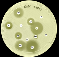
Step-by-Step Guidance
Observe the plate for clear zones around each antibiotic disk.
Compare the size of the zones to determine sensitivity or resistance.
Remember that a larger zone does not always mean greater effectiveness—consider molecular weight and diffusion rate.
Try solving on your own before revealing the answer!
Q14. What dye in EMB inhibits the growth of Gram-positive organisms?
Background
Topic: Eosin Methylene Blue (EMB) Agar
This question tests your knowledge of selective agents in differential media.
Key Terms and Concepts:
Dyes: Eosin Y and methylene blue inhibit Gram-positive bacteria.
Step-by-Step Guidance
Recall the purpose of EMB agar and which dyes are present.
Explain how these dyes function as selective agents.
Try solving on your own before revealing the answer!
Q15. What type of hemolysis does S. pyogenes cause on blood agar?
Background
Topic: Hemolytic Patterns of Streptococcus Species
This question tests your ability to associate specific organisms with their hemolytic activity.
Key Terms and Concepts:
Beta Hemolysis: Complete clearing of red blood cells around colonies.
Step-by-Step Guidance
Recall the hemolytic pattern produced by S. pyogenes.
Describe how this appears on blood agar.
Try solving on your own before revealing the answer!
Q16. What causes the hazy green color in alpha hemolysis?
Background
Topic: Alpha Hemolysis Mechanism
This question tests your understanding of the biochemical basis for alpha hemolysis.
Key Terms and Concepts:
Hydrogen Peroxide: Produced by some bacteria, oxidizes hemoglobin to methemoglobin, causing greenish color.
Step-by-Step Guidance
Recall the metabolic byproducts of alpha-hemolytic bacteria.
Explain how these byproducts interact with red blood cells to produce the observed color.
Try solving on your own before revealing the answer!
Q17. What are the differences between antisepsis, degerming, sterilization, sanitization, and disinfection?
Background
Topic: Microbial Control Terminology
This question tests your ability to distinguish between different methods of microbial control and their applications.
Key Terms and Concepts:
Antisepsis: Use of chemicals on living tissue to reduce microbes.
Degerming: Mechanical removal of microbes from living tissue (e.g., handwashing).
Sterilization: Complete destruction of all microbes, including spores.
Sanitization: Reducing microbes to safe levels on inanimate objects.
Disinfection: Reducing or eliminating most microbes on inanimate objects (not spores).
Step-by-Step Guidance
Define each term and give an example of its application.
Compare and contrast the effectiveness and appropriate use of each method.
Try solving on your own before revealing the answer!
Q18. How do length of exposure and amount of bacteria affect germicide efficacy?
Background
Topic: Factors Affecting Germicide Effectiveness
This question tests your understanding of how microbial load and exposure time influence the success of disinfection or sterilization.
Key Terms and Concepts:
Length of Exposure: Longer exposure increases effectiveness.
Microbial Load: More bacteria require longer or stronger treatment.
Step-by-Step Guidance
Explain why a higher number of bacteria or biofilm requires longer exposure to germicides.
Describe how these factors are considered in laboratory and clinical protocols.
Try solving on your own before revealing the answer!
Q19. What types of methods are considered safe for use on human tissues?
Background
Topic: Safe Microbial Control Methods
This question tests your knowledge of which antimicrobial methods are appropriate for living tissues.
Key Terms and Concepts:
Antiseptics: Chemicals safe for use on skin or mucous membranes.
Step-by-Step Guidance
List examples of antiseptics and their uses.
Explain why other methods (e.g., disinfectants, sterilants) are not suitable for living tissue.
Try solving on your own before revealing the answer!
