 Back
Microbiology Review: Metabolism, Growth, Pathogens, and Eukaryotic Microbes
Study Guide - Smart Notes
 Tailored notes based on your materials, expanded with key definitions, examples, and context.
Q1. Fill in the following table for Glycolysis, Transition Step, Krebs Cycle, and Electron Transport Chain:
Background
Topic: Microbial Metabolism
This question tests your understanding of the major metabolic pathways in cellular respiration, including the substrates, products, and key steps of glycolysis, the transition step, the Krebs cycle, and the electron transport chain.
Key Terms and Formulas
Glycolysis: The breakdown of glucose to pyruvate, producing ATP and NADH.
Transition Step: Conversion of pyruvate to acetyl-CoA, producing NADH and CO2.
Krebs Cycle: Series of reactions that generate ATP, NADH, FADH2, and CO2 from acetyl-CoA.
Electron Transport Chain (ETC): Uses electrons from NADH and FADH2 to create a proton gradient for ATP synthesis.
Substrate-Level Phosphorylation (SLP): Direct formation of ATP in metabolic pathways.
Step-by-Step Guidance
For each process (glycolysis, transition step, Krebs cycle, ETC), identify the starting substrate and the main end product(s).
List the key products/byproducts for each process, such as ATP (via SLP), NADH, FADH2, and CO2.
For the ETC, recall when electrons are pumped and the role of the proton motive force and chemiosmosis.
Remember the ATP yield per NADH and FADH2 in the ETC.
Fill in the table with the correct substrates, products, and counts for each process, using your knowledge of cellular respiration.
Try solving on your own before revealing the answer!
Final Answer:
Glycolysis:
First substrate: Glucose
End product: Pyruvate
1) ATP (SLP): 2 net
2) CO2: 0
3) NADH: 2
Transition Step:
Enzyme: Pyruvate dehydrogenase
End product: Acetyl-CoA
1) NADH: 2 (per glucose)
2) CO2: 2 (per glucose)
Krebs Cycle:
First substrate: Acetyl-CoA
1) ATP (SLP): 2 (per glucose)
2) CO2: 4 (per glucose)
3) NADH: 6 (per glucose)
4) FADH2: 2 (per glucose)
Electron Transport Chain:
Electrons are pumped as NADH and FADH2 donate electrons to the chain.
Proton motive force: Electrochemical gradient of protons across the membrane.
Chemiosmosis: ATP synthesis driven by the flow of protons back through ATP synthase.
ATP per NADH: 2.5–3
ATP per FADH2: 1.5–2
Q2. The diffusion of water across a semipermeable membrane is driven by unequal solute concentrations on the two sides of a biological membrane. What is this process called?
Background
Topic: Membrane Transport
This question tests your knowledge of passive transport mechanisms in cells, specifically the movement of water.
Key Terms
Osmosis: The diffusion of water across a semipermeable membrane due to differences in solute concentration.
Semipermeable membrane: A barrier that allows certain molecules (like water) to pass but not others (like solutes).
Step-by-Step Guidance
Recall the term for water movement across membranes in response to solute concentration differences.
Think about how this process is essential for maintaining cell homeostasis.
Write the term that describes this process.
Try solving on your own before revealing the answer!
Final Answer: Osmosis
Osmosis is the process by which water diffuses across a semipermeable membrane from an area of low solute concentration to high solute concentration.
Q3. This kind of pressure may have dire effects on a cell. Give at least one example of how a bacterial cell wall affords some protection.
Background
Topic: Cell Structure and Function
This question is about the role of the bacterial cell wall in protecting cells from osmotic pressure.
Key Terms
Osmotic pressure: The pressure exerted by water moving across a membrane due to osmosis.
Cell wall: A rigid structure that surrounds the cell membrane in bacteria, providing structural support and protection.
Step-by-Step Guidance
Consider what happens to cells in hypotonic environments (where water enters the cell).
Think about the function of the bacterial cell wall in preventing cell lysis.
Describe one way the cell wall helps bacteria survive osmotic stress.
Try solving on your own before revealing the answer!
Final Answer:
The bacterial cell wall prevents the cell from bursting (lysis) in hypotonic environments by providing structural support and counteracting osmotic pressure.
Q4. 70% of bacterial infections such as kidney infections, gum decay, and health care associated infections are caused by _________. Quorum sensing is a process used by microbes to respond to the density of microbes nearby. Describe the development of this microbial community.
Background
Topic: Microbial Communities and Communication
This question tests your understanding of biofilms and quorum sensing in bacteria.
Key Terms
Biofilm: A complex community of microorganisms attached to a surface and embedded in a self-produced matrix.
Quorum sensing: Cell-to-cell communication mechanism that allows bacteria to coordinate gene expression based on population density.
Step-by-Step Guidance
Recall the term for structured microbial communities that cause persistent infections.
Think about how bacteria use chemical signals to sense their population density.
Describe the steps in biofilm development, including attachment, growth, and maturation.
Try solving on your own before revealing the answer!
Final Answer:
Biofilms. Bacteria attach to a surface, produce extracellular polymeric substances, communicate via quorum sensing, and form a mature, structured community resistant to antibiotics and immune responses.
Q5. A generation time or doubling time is the time required for a bacterial cell to grow and divide. Staph aureus has a generation time of 20 minutes or less. If 50 cells are placed in a broth, and the lag time is one generation time, how many hours will it take for the broth to have over 12,000 bacteria?
Background
Topic: Microbial Growth
This question tests your ability to calculate bacterial population growth using generation time and exponential growth equations.
Key Formula
Where = final number of cells, = initial number of cells, = number of generations.
Generation time (): Time required for one cell division.
Step-by-Step Guidance
Identify the initial number of cells () and the target number ().
Calculate the number of generations needed using the formula .
Use logarithms to solve for .
Multiply the number of generations by the generation time (20 minutes) to get the total time.
Convert the total time from minutes to hours.
Try solving on your own before revealing the answer!
Final Answer:
It will take approximately 3.3 hours for the population to exceed 12,000 bacteria.
Calculation: generations; minutes minutes hours plus 20 minutes lag time hours, rounded up to the next full generation.
Q6. Name the phase on a typical microbial growth curve:
1) When the number of dying cells = number of cells being produced.
2) When bacterial cells are most vulnerable to antimicrobial drugs that interfere with metabolism.
3) When the number of dying cells exceeds the number of cells being produced.
4) When cells are adjusting to a new environment.
Background
Topic: Microbial Growth Phases
This question tests your knowledge of the phases of the bacterial growth curve and their characteristics.
Key Terms
Lag phase: Adjustment period after inoculation.
Log (exponential) phase: Rapid cell division and growth.
Stationary phase: Rate of cell growth equals rate of cell death.
Death (decline) phase: Cell death rate exceeds cell growth rate.
Step-by-Step Guidance
Match each description to the correct phase of the growth curve.
Recall the order and characteristics of each phase.
Write the name of the phase for each description.
Try solving on your own before revealing the answer!
Final Answer:
1) Stationary phase
2) Log (exponential) phase
3) Death (decline) phase
4) Lag phase
Q7. Circle the forms of oxygen that steal electrons from other molecules: molecular oxygen, oxygen in an organic molecule, singlet oxygen, peroxide anion, superoxide radicals
Background
Topic: Reactive Oxygen Species (ROS)
This question tests your knowledge of which forms of oxygen are reactive and can cause oxidative damage in cells.
Key Terms
Reactive Oxygen Species (ROS): Highly reactive forms of oxygen that can damage cellular components.
Examples: Singlet oxygen, superoxide radicals, peroxide anion.
Step-by-Step Guidance
Identify which forms of oxygen are considered ROS and can steal electrons (oxidize other molecules).
Recall that molecular oxygen and oxygen in organic molecules are generally not reactive in this way.
Circle the correct forms.
Try solving on your own before revealing the answer!
Final Answer:
Singlet oxygen, peroxide anion, and superoxide radicals are the forms that steal electrons from other molecules.
Q8. Stage the mitosis phase and give the number of chromosomes for each example. All cells depicted are diploid:
Background
Topic: Eukaryotic Cell Division
This question tests your understanding of the stages of mitosis and chromosome number in diploid cells.
Key Terms
Mitosis: Process of eukaryotic cell division resulting in two identical daughter cells.
Diploid: Cells with two sets of chromosomes (2n).
Stages: Prophase, metaphase, anaphase, telophase.
Step-by-Step Guidance
Identify the mitosis stage based on chromosome arrangement and cell structure.
Recall the chromosome number for diploid cells at each stage.
Write the stage and chromosome number for each example.
Try solving on your own before revealing the answer!
Final Answer:
Answers will depend on the specific examples provided, but for diploid cells (2n):
Prophase: 2n chromosomes
Metaphase: 2n chromosomes aligned at the metaphase plate
Anaphase: Sister chromatids separate, still 2n but now counted as individual chromosomes
Telophase: 2n chromosomes in each new nucleus
Q9. Identify the following protozoans:
A parabasalid that lives in the human vagina and when the pH is raised may proliferate and cause severe inflammation.
A Diplomonad that has two equal sized nuclei and multiple flagella, may form cysts and is a diarrhea causing pathogen.
A kinetoplastid that has species that cause African sleeping sickness or Chaga’s disease.
An apicomplexan that causes malaria.
An apicomplexan that causes toxoplasmosis.
An apicomplexan that causes cryptosporidiosis.
The subgroup of Alveolates that make up plankton, may be bioluminescent, may produce neurotoxins and has two flagella.
Background
Topic: Medically Important Protozoa
This question tests your ability to identify protozoan pathogens and their characteristics.
Key Terms
Parabasalid: Trichomonas vaginalis
Diplomonad: Giardia lamblia
Kinetoplastid: Trypanosoma species
Apicomplexans: Plasmodium (malaria), Toxoplasma (toxoplasmosis), Cryptosporidium (cryptosporidiosis)
Dinoflagellates: Alveolates with two flagella, some bioluminescent, some produce toxins
Step-by-Step Guidance
Match each description to the correct protozoan genus or group.
Recall the diseases and characteristics associated with each group.
Write the name of the protozoan for each description.
Try solving on your own before revealing the answer!
Final Answer:
Trichomonas vaginalis
Giardia lamblia
Trypanosoma species
Plasmodium species
Toxoplasma gondii
Cryptosporidium species
Dinoflagellates
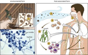
Q10. Histoplasma capsulatum: What fungal characteristic is being depicted in the image below?
Background
Topic: Fungal Pathogens and Morphology
This question tests your ability to recognize fungal dimorphism and environmental/host-associated forms.
Key Terms
Dimorphism: The ability of some fungi to exist in two different forms depending on environmental conditions (e.g., mold in the environment, yeast in the host).
Conidia: Asexual spores produced by fungi.
Hyphae: Filamentous structures of molds.
Step-by-Step Guidance
Examine the image for evidence of two forms: environmental (mold) and host-associated (yeast).
Identify the structures labeled as conidia and hyphae in the environmental form.
Note the transition to yeast-like cells in the host-associated form.
Recall the term for this ability to switch forms.
Try solving on your own before revealing the answer!
Final Answer:
The image depicts fungal dimorphism: Histoplasma capsulatum exists as a mold (with hyphae and conidia) in the environment and as a yeast in host tissues.
Q11. Distinguish between hyphae and mycelium.
Background
Topic: Fungal Structure
This question tests your understanding of the basic structural components of fungi.
Key Terms
Hyphae: Long, thread-like filaments that make up the body of a fungus.
Mycelium: A mass of hyphae that forms the vegetative part of a fungus.
Step-by-Step Guidance
Define hyphae and describe their appearance and function.
Define mycelium and explain how it relates to hyphae.
Compare and contrast the two terms.
Try solving on your own before revealing the answer!
Final Answer:
Hyphae are individual filamentous structures, while mycelium is the collective mass of hyphae that forms the main body of a fungus.
Q12. Distinguish between a biofilm and lichens.
Background
Topic: Microbial Communities
This question tests your understanding of different types of microbial associations.
Key Terms
Biofilm: A community of microorganisms attached to a surface and embedded in a self-produced matrix.
Lichen: A symbiotic association between a fungus and a photosynthetic partner (alga or cyanobacterium).
Step-by-Step Guidance
Define biofilm and describe its structure and function.
Define lichen and explain the relationship between its components.
Compare the two in terms of composition and ecological role.
Try solving on your own before revealing the answer!
Final Answer:
Biofilms are communities of bacteria and other microbes attached to surfaces, while lichens are symbiotic associations between fungi and photosynthetic organisms.
Q13. Name the eukaryote that acts as a vector for each of the following diseases: Malaria, Rocky mountain spotted fever, Chaga’s disease, African sleeping sickness, Lyme disease, Typhus, Leishmania
Background
Topic: Disease Vectors
This question tests your knowledge of the eukaryotic organisms that transmit various infectious diseases.
Key Terms
Vector: An organism that transmits a pathogen from one host to another.
Examples: Mosquitoes, ticks, sandflies, etc.
Step-by-Step Guidance
Recall the vector for each disease listed.
Match the disease to the correct eukaryotic vector (e.g., mosquito, tick, sandfly).
Write the name of the vector for each disease.
Try solving on your own before revealing the answer!
Final Answer:
Malaria: Anopheles mosquito
Rocky mountain spotted fever: Tick (Dermacentor species)
Chaga’s disease: Reduviid (kissing) bug
African sleeping sickness: Tsetse fly
Lyme disease: Ixodes tick
Typhus: Louse (Pediculus humanus)
Leishmania: Sandfly
