 Back
BackMicrobiology: Skin and Soft Tissue Infections – Study Guidance
Study Guide - Smart Notes
 Tailored notes based on your materials, expanded with key definitions, examples, and context.
Q1. Moisture in perspiration encourages microbial growth. What perspiration factors discourage growth?
Background
Topic: Skin Microbiology & Host Defenses
This question tests your understanding of the skin's natural defenses against microbial colonization, especially in the context of perspiration.
Key Terms and Concepts:
Perspiration: Fluid secreted by sweat glands, containing water, salts, and other substances.
Antimicrobial factors: Substances or conditions that inhibit microbial growth.
Step-by-Step Guidance
Recall that while moisture provides a suitable environment for microbes, sweat also contains substances that can inhibit their growth.
Think about the chemical composition of sweat—what antimicrobial molecules are present?
Consider the pH of the skin and how it affects microbial survival.
List at least two factors in perspiration that actively discourage microbial growth.
Try solving on your own before revealing the answer!
Final Answer:
Factors in perspiration that discourage microbial growth include:
High salt concentration (hypertonic environment) inhibits many microbes.
Lysozyme, an enzyme in sweat, breaks down bacterial cell walls.
Fatty acids and low pH (acidic environment) also inhibit microbial growth.
These factors create a hostile environment for many pathogens, helping protect the skin.
Q2. Are your normal skin bacteria more likely to be gram-positive or gram-negative? Why?
Background
Topic: Normal Microbiota of the Skin
This question tests your knowledge of the types of bacteria that commonly inhabit human skin and the reasons for their predominance.
Key Terms:
Gram-positive bacteria: Bacteria with thick peptidoglycan cell walls, stain purple in Gram stain.
Gram-negative bacteria: Bacteria with thin peptidoglycan and outer membrane, stain pink/red.
Step-by-Step Guidance
Recall the environmental conditions of the skin (dry, salty, acidic).
Think about which bacterial cell wall structure is more resistant to these harsh conditions.
Consider examples of common skin bacteria and their Gram reaction.
Explain why one group is more prevalent than the other on the skin.
Try solving on your own before revealing the answer!
Final Answer:
Normal skin bacteria are more likely to be gram-positive. This is because gram-positive bacteria, such as Staphylococcus and Micrococcus species, have thick peptidoglycan cell walls that help them resist desiccation and high salt concentrations found on the skin. Gram-negative bacteria are less tolerant of these conditions.
Q3. Compare and contrast the following terms: a) Exanthem vs. enanthem b) vesicle, macule, papule, pustule
Background
Topic: Skin Lesions and Rashes
This question assesses your ability to distinguish between different types of skin and mucous membrane lesions, which is important for clinical diagnosis in microbiology.
Key Terms:
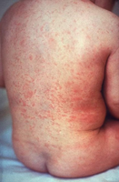
Exanthem: Widespread rash on the skin.
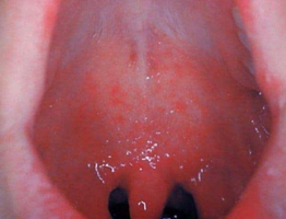
Enanthem: Rash on mucous membranes.
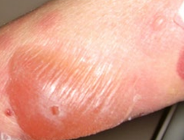
Vesicle: Small, fluid-filled blister.
Macule: Flat, discolored spot.
Papule: Raised, solid lesion.
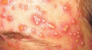
Pustule: Raised lesion filled with pus.
Step-by-Step Guidance
Define exanthem and enanthem, focusing on their locations.
List the differences in appearance and clinical significance between the two.
Define vesicle, macule, papule, and pustule, noting their physical characteristics (elevation, contents, color).
Compare and contrast these lesion types in terms of their appearance and what they indicate about underlying pathology.
Try solving on your own before revealing the answer!
Final Answer:
a) Exanthem is a rash that appears on the skin, while enanthem is a rash that appears on mucous membranes (such as inside the mouth).
b) Vesicle: small, fluid-filled blister; Macule: flat, discolored spot; Papule: raised, solid lesion; Pustule: raised lesion filled with pus. These terms help describe the appearance and possible cause of skin lesions.
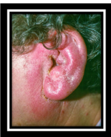
Q4. What is the colloquial name for otitis externa? What is the most common causative agent?
Background
Topic: Bacterial Infections of the Ear
This question tests your knowledge of common ear infections, their names, and the typical pathogens involved.
Key Terms:
Otitis externa: Infection of the outer ear canal.
Colloquial name: Common or layperson's term for a disease.
Causative agent: The microorganism responsible for the infection.
Step-by-Step Guidance
Recall the common name for otitis externa, especially in the context of swimming or water exposure.
Think about the environmental conditions that favor the growth of the most common causative agent.
Identify the typical bacterium associated with this infection.
Try solving on your own before revealing the answer!
Final Answer:
The colloquial name for otitis externa is "swimmer's ear." The most common causative agent is Pseudomonas aeruginosa, a bacterium that thrives in moist environments like swimming pools.
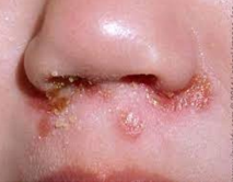
Q5. A 4-year-old girl comes to your clinic with a rash around her nose that includes a few small pustules and yellow scabs. Microscopic and biochemical examination of the bacteria cultured from the lesions show catalase-negative, Gram-positive cocci, that look like a string of pearls. What condition does this girl have and what is the causative agent?
Background
Topic: Bacterial Skin Infections – Impetigo
This question tests your ability to interpret clinical and laboratory findings to diagnose a common pediatric skin infection and identify the causative organism.
Key Terms:
Catalase-negative: Does not produce the enzyme catalase.
Gram-positive cocci: Spherical bacteria that retain the crystal violet stain.
Impetigo: Superficial skin infection, often with honey-colored crusts.
Step-by-Step Guidance
Analyze the clinical presentation: rash, pustules, yellow scabs around the nose.
Interpret the lab findings: catalase-negative, Gram-positive cocci in chains.
Recall which bacteria fit this description and are common causes of impetigo.
Identify the condition and the most likely causative agent based on the evidence.
Try solving on your own before revealing the answer!
Final Answer:
The girl has impetigo. The causative agent is Streptococcus pyogenes (Group A Streptococcus), which is catalase-negative and forms chains of Gram-positive cocci.
Q6. You have a pediatric patient with SSSS. What does that stand for? Microbiological culture of the blister fluid comes back negative for any organisms. Explain why. How would you treat this patient?
Background
Topic: Staphylococcal Skin Syndromes
This question tests your understanding of Staphylococcal Scalded Skin Syndrome (SSSS), its pathogenesis, and appropriate treatment strategies.
Key Terms:
SSSS: Staphylococcal Scalded Skin Syndrome
Exotoxin: Toxin secreted by bacteria that can cause tissue damage.
Culture-negative: No bacteria detected in the sample.
Step-by-Step Guidance
Define what SSSS stands for and describe its main clinical features.
Explain why the blister fluid might be sterile (no organisms found on culture).
Discuss the role of bacterial toxins in this syndrome.
Outline the general principles of treatment for SSSS.

Try solving on your own before revealing the answer!
Final Answer:
SSSS stands for Staphylococcal Scalded Skin Syndrome. The blister fluid is culture-negative because the disease is caused by exfoliative toxins produced by Staphylococcus aureus, not by the direct presence of bacteria in the blisters. Treatment involves antibiotics effective against S. aureus and supportive care for skin healing and fluid balance.
Q7. Why do some strains of S. pyogenes cause “flesh-eating” disease, while others do not?
Background
Topic: Streptococcal Virulence Factors
This question tests your understanding of the genetic and molecular basis for differences in pathogenicity among strains of Streptococcus pyogenes.
Key Terms:
Necrotizing fasciitis: Severe, rapidly spreading soft tissue infection (“flesh-eating” disease).
Virulence factors: Molecules produced by pathogens that contribute to their ability to cause disease.
Step-by-Step Guidance
Recall what “flesh-eating” disease refers to and which organism is involved.
Think about the role of bacterial toxins and enzymes in tissue destruction.
Consider genetic differences between strains that might explain varying severity.
List at least two virulence factors that are important for necrotizing fasciitis.
Try solving on your own before revealing the answer!
Final Answer:
Some strains of S. pyogenes produce additional virulence factors, such as streptococcal pyrogenic exotoxins and enzymes like streptokinase and hyaluronidase, which allow them to invade and destroy deeper tissues, leading to necrotizing fasciitis. Other strains lack these factors and cause milder infections.
Q8. Explain the coagulase test. What bacteria are identified by a positive coagulase test? What bacteria are characterized as group A beta-hemolytic?
Background
Topic: Bacterial Identification – Staphylococcus and Streptococcus
This question tests your knowledge of laboratory tests used to differentiate between important bacterial pathogens.
Key Terms and Formulas:
Coagulase test: Detects the enzyme coagulase, which causes blood plasma to clot.
Beta-hemolysis: Complete lysis of red blood cells on blood agar.
Group A Streptococcus: Streptococcus pyogenes
Step-by-Step Guidance
Describe how the coagulase test is performed and what a positive result looks like.
Identify which common skin pathogen is coagulase-positive.
Explain what "group A beta-hemolytic" means and which bacterium it refers to.
Try solving on your own before revealing the answer!
Final Answer:
The coagulase test detects the enzyme coagulase, which causes plasma to clot. Staphylococcus aureus is identified by a positive coagulase test. Group A beta-hemolytic bacteria are Streptococcus pyogenes.
Q9. A laboratory test used to determine the identity of Staphylococcus aureus is its growth on mannitol salt agar. The medium contains 7.5% sodium chloride (NaCl). Why is it considered a selective medium for S. aureus?
Background
Topic: Selective and Differential Media
This question tests your understanding of how certain media are used to isolate and identify specific bacteria based on their growth characteristics.
Key Terms:
Mannitol salt agar: A medium with high salt concentration and mannitol sugar.
Selective medium: Inhibits growth of some organisms while allowing others to grow.
Step-by-Step Guidance
Recall the salt tolerance of Staphylococcus aureus compared to other bacteria.
Explain how the high salt concentration affects the growth of different bacteria.
Describe why this property makes mannitol salt agar selective for S. aureus.
Try solving on your own before revealing the answer!
Final Answer:
Mannitol salt agar is selective for S. aureus because its high salt concentration (7.5% NaCl) inhibits the growth of most other bacteria, but S. aureus can tolerate and grow in these conditions.
Q10. Analyses of nine conjunctivitis cases provided the data in the table below. How were these infections transmitted? How could they be prevented?
Background
Topic: Transmission and Prevention of Eye Infections
This question tests your ability to interpret epidemiological data and apply infection control principles.
Key Terms:
Conjunctivitis: Inflammation of the conjunctiva (pink eye).
Transmission: How the infection spreads from person to person.
Prevention: Measures to stop the spread of infection.
Step-by-Step Guidance
Review the data for patterns (e.g., common exposures, contacts, or behaviors).
Identify likely modes of transmission (direct contact, fomites, etc.).
List at least two strategies for preventing the spread of conjunctivitis.
Try solving on your own before revealing the answer!
Final Answer:
The conjunctivitis infections were likely transmitted by direct contact or contaminated objects (fomites). Prevention includes good hand hygiene, avoiding sharing towels or personal items, and disinfecting surfaces.
Q11. A hospitalized patient recovering from surgery develops an infection that has blue-green pus and a grapelike odor. What is the probable etiology? How might the patient have acquired this infection?
Background
Topic: Nosocomial Infections – Pseudomonas
This question tests your ability to recognize classic clinical features of Pseudomonas aeruginosa infections and understand hospital-acquired infection risks.
Key Terms:
Nosocomial infection: Hospital-acquired infection.
Pseudomonas aeruginosa: Gram-negative bacterium known for blue-green pigment and fruity odor.
Step-by-Step Guidance
Identify the organism based on the description of pus color and odor.
Consider common sources of this bacterium in hospital settings.
Explain how a surgical patient might become infected.
Try solving on your own before revealing the answer!
Final Answer:
The probable etiology is Pseudomonas aeruginosa. The patient may have acquired the infection from contaminated hospital equipment, surfaces, or solutions, especially if they had a wound or compromised immune system.
Q12. A 12-year-old diabetic girl using continuous subcutaneous insulin infusion to manage her diabetes developed a fever (39.4°C), low blood pressure, abdominal pain, and erythroderma. She was supposed to change the needle-insertion site every 3 days after cleaning the skin with an iodine solution. Frequently she did not change the insertion site more often than every 10 days. Blood culture was negative, and abscesses at insertion sites were not cultured. What is the probable cause of her symptoms?
Background
Topic: Device-Associated Infections and Toxic Shock
This question tests your ability to connect clinical presentation and risk factors to a likely diagnosis, especially in the context of toxin-mediated disease.
Key Terms:
Erythroderma: Widespread redness of the skin.
Toxin-mediated disease: Illness caused by bacterial toxins rather than direct infection.
Step-by-Step Guidance
Analyze the risk factors (diabetes, infrequent site changes, possible skin colonization).
Consider the symptoms (fever, hypotension, erythroderma) and what syndrome they suggest.
Explain why blood cultures might be negative in this scenario.
Identify the most likely causative organism and syndrome.
Try solving on your own before revealing the answer!
Final Answer:
The probable cause is toxic shock syndrome due to Staphylococcus aureus toxin production at the insulin infusion site. The blood culture is negative because the symptoms are caused by toxins, not by bacteria in the bloodstream.
Q13. A teenaged male with confirmed influenza was hospitalized when he developed respiratory distress. He had a fever, rash, and low blood pressure. S. aureus was isolated from his respiratory secretions. Discuss the relationship between his symptoms and the etiological agent.
Background
Topic: Secondary Bacterial Infections and Toxin-Mediated Disease
This question tests your understanding of how viral infections can predispose to severe bacterial complications, including toxin-mediated syndromes.
Key Terms:
Secondary infection: Bacterial infection following a viral illness.
Toxin-mediated syndrome: Disease caused by bacterial toxins (e.g., toxic shock).
Step-by-Step Guidance
Consider how influenza can damage respiratory defenses and allow bacterial superinfection.
Relate the symptoms (fever, rash, hypotension) to possible toxin-mediated effects.
Explain the role of S. aureus in causing these symptoms after influenza.
Try solving on your own before revealing the answer!
Final Answer:
The patient's influenza likely damaged his respiratory tract, allowing S. aureus to infect and produce toxins. The combination of fever, rash, and low blood pressure suggests toxic shock syndrome, a toxin-mediated complication of S. aureus infection.
