 Back
Microbiology Study Guide: Cell Structure, Laboratory Tools, and Bacterial Diseases
Study Guide - Smart Notes
 Tailored notes based on your materials, expanded with key definitions, examples, and context.
Introduction to Microbiology
Microbes in Our Lives
Microorganisms are organisms too small to be seen with the unaided eye. While only a few are pathogenic (disease-causing), microbes play essential roles in decomposing organic waste, producing industrial chemicals (such as ethanol and acetone), fermenting foods, and creating products used in disease treatment.
Microorganisms: Include bacteria, fungi, protozoa, helminths, viruses, and prions.
Applications: Industrial, environmental, and medical.
Microbial Structure
Microbes are classified into three main groups based on cellular structure:
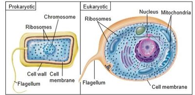
Prokaryotes: Microscopic unicellular organisms lacking nuclei and membrane-bound organelles (e.g., bacteria).
Eukaryotes: Unicellular or multicellular organisms with nuclei and membrane-bound organelles (e.g., fungi, protozoa, helminths).
Acellular microbes: Viruses and prions, which are parasitic particles.
Classifying Organisms
Taxonomy is the science of organizing, classifying, and naming living things. The formal system originated by Carl Linnaeus includes:
Classification: Orderly arrangement into groups.
Identification: Determining traits for placement in taxonomic schemes.
Nomenclature: Assigning names.
Taxonomic hierarchy ranks: Domain, Kingdom, Phylum, Class, Order, Family, Genus, Species.
Naming Microorganisms
Each organism has a genus and specific epithet (e.g., Staphylococcus aureus).
Scientific names are italicized or underlined; genus is capitalized, specific epithet is lowercase.
Abbreviations: E. coli (large intestine), S. aureus (skin).
Tools of the Laboratory
The Five I's of Microbiology
Microbiologists use five main steps to study microorganisms:
Inoculation: Introducing a sample into a sterile medium for growth.
Incubation: Growing the inoculum under controlled conditions.
Isolation: Separating one species from another, often using the streak plate method.
Inspection: Observing colonies macroscopically and cells microscopically.
Identification: Determining the species and characteristics of the microbe.
Methods of Culturing Microorganisms
Culture medium: Nutrients prepared for microbial growth.
Inoculum: Microbe introduced to the medium.
Aseptic technique: Prevents contamination by unwanted organisms.
Types of Media
General purpose media: Supports growth of many microbes (e.g., nutrient agar).
Enriched media: Supports fastidious bacteria.
Selective media: Suppresses unwanted microbes, encourages desired ones (e.g., mannitol salt agar).
Differential media: Distinguishes colonies of different microbes.
Maintenance & Disposal of Cultures
Autoclave and incineration are used for sterilization and disposal.
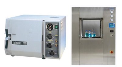
Microscopes
Key Characteristics
Microscopes are essential for visualizing microorganisms. Their reliability depends on:
Magnification: Ability to enlarge objects.
Resolution: Ability to distinguish two adjacent objects.
Refractive index: Measure of light-bending ability; immersion oil is used to prevent light loss at high magnification.
Types of Microscopes
Light microscope: Allows observation of live specimens; limited resolution.
Electron microscope: High resolution and depth of field; specimens must be killed and coated with metals.
Preparing Specimens for Light Microscopes
Wet mounts and hanging drop mounts: Examine live cells for motility, shape, and arrangement.
Fixed mounts: Dried and heated films stained with dyes for visualization.
Staining Techniques
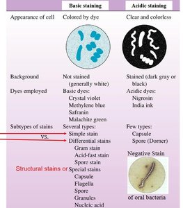
Basic stains: Positively charged dyes (e.g., crystal violet, safranin).
Acidic stains: Negatively charged dyes (e.g., nigrosin, India ink).
Negative staining: Stains background, not cells; useful for accurate size and spirochetes.
Simple vs. Differential Staining
Simple stains: Use one dye.
Differential stains: Use two dyes to distinguish cell types or structures (e.g., Gram stain, endospore stain, acid-fast stain).
Gram Stain
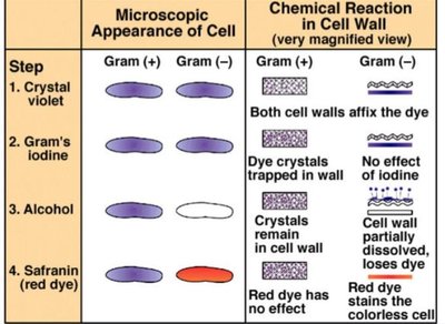
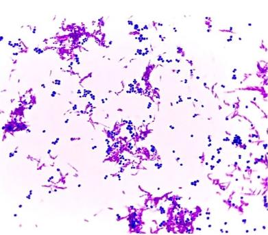
The Gram stain is a differential stain that distinguishes between Gram-positive and Gram-negative bacteria based on cell wall structure.
Gram-positive: Retain crystal violet, stain purple.
Gram-negative: Lose crystal violet, stain red with safranin.
Endospore Stain
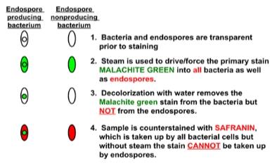
Endospore staining differentiates endospore-producing bacteria from non-producing ones using malachite green and safranin.
Prokaryotic Cell Structures & Functions
Characteristics of Cells & Life
All living cells share basic features: shape, internal content (cytoplasm), DNA, ribosomes, and metabolic capabilities. Prokaryotic cells (bacteria and archaea) lack a nucleus and membrane-bound organelles, while eukaryotic cells (animals, plants, fungi, protists) have these structures.
Bacterial Shapes, Arrangements, and Sizes
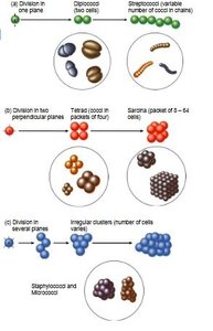
Coccus: Spherical
Bacillus: Rod-shaped
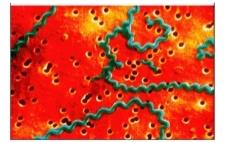
Spirillum: Spiral-shaped
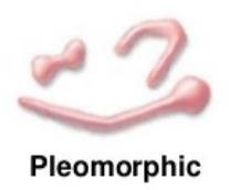
Pleomorphism: Variation in shape and size within a species, often due to environmental conditions.
Prokaryotic Cell Structure
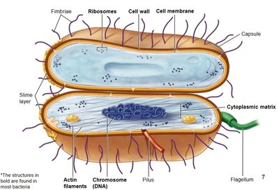
Bacterial cells have several key structures:
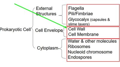
External structures: Glycocalyx, flagella, fimbriae, pili
Cell envelope: Cell wall, cell membrane
Cytoplasm: Water, ribosomes, nucleoid chromosome, plasmids, endospores
External Structures
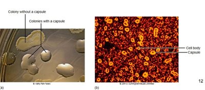
Glycocalyx: Surface coating made of sugars and/or proteins; includes slime layer (loose) and capsule (tight).
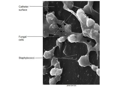
Functions: Protection from dehydration, nutrient loss, phagocytosis, and attachment (biofilm formation).
Flagella
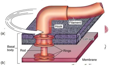
Structure: Filament, hook, basal body
Function: Motility; rotates 360°
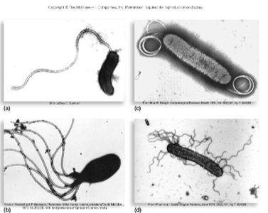
Arrangements: Monotrichous (single), lophotrichous (cluster), amphitrichous (both ends), peritrichous (all over)
Fimbriae and Pili
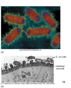
Fimbriae: Hairlike bristles for adhesion
Pili: Tubular structures for DNA transfer (conjugation); found only in Gram-negative cells
Cell Envelope
Cell wall: Determines shape, prevents lysis; primary component is peptidoglycan
Gram-positive: Thick peptidoglycan, teichoic acids
Gram-negative: Thin peptidoglycan, outer membrane with lipopolysaccharides (LPS), porin proteins
Nontypical cell walls: Mycobacterium (mycolic acid), mycoplasma (no cell wall, stabilized by sterols)
Cell Membrane
Phospholipid bilayer with embedded proteins (fluid mosaic model)
Functions: Energy reactions, nutrient processing, selective permeability
Internal Structures
Cytoplasm: Gelatinous solution, 70-80% water
Nucleoid: Single circular double-stranded DNA chromosome
Plasmids: Small, circular DNA; not essential for growth, used in genetic engineering
Ribosomes: Site of protein synthesis; made of rRNA and protein
Endospores: Highly resistant, dormant structures produced by some Gram-positive genera (e.g., Clostridium, Bacillus)
Microbial Infections: Skin, Nervous, Respiratory, Digestive, and Urogenital Systems
Necrotizing Fasciitis (Skin)
Necrotizing fasciitis is a severe bacterial infection of the skin, most commonly caused by Streptococcus pyogenes, but also by Staphylococcus aureus and Clostridium perfringens.
Symptoms: Intense pain, swelling, rapid tissue destruction, necrosis
Transmission: Direct contact, entry through wounds
Treatment: Aggressive surgical removal, broad-spectrum antibiotics
Bacterial Meningitis (Nervous System)
Bacterial meningitis is an infection of the meninges and cerebrospinal fluid, most often caused by Streptococcus pneumoniae, Neisseria meningitidis, Haemophilus influenzae, Listeria monocytogenes, and Streptococcus agalactiae.
Symptoms: High fever, meningeal inflammation, photophobia, headache, stiff neck, nausea, vomiting, neurological damage
Diagnosis: Symptoms, spinal tap, Gram stain, culture
Treatment: Immediate IV antibiotics (cephalosporins, vancomycin)
Prevention: Vaccination, prophylactic treatment for close contacts
Tuberculosis (Respiratory System)
Tuberculosis is a bacterial infection of the respiratory tract caused by Mycobacterium tuberculosis. Transmission occurs via droplets.
Symptoms: Breathing difficulty, fatigue, weight loss, chest pain, coughing up blood
Treatment: Long-term multi-drug therapy, BCG vaccine
Peptic Ulcers (Digestive System)
Peptic ulcers are erosions of the GI tract lining, caused by Helicobacter pylori.
Symptoms: Abdominal pain, nausea, vomiting, weight loss, bloody stool
Diagnosis: Biopsy, Gram stain, urease test, urea breath test
Treatment: Antibiotics, acid inhibitors
Prevention: Reduce risk factors (NSAIDs, alcohol, smoking, stress)
Chlamydia (Urogenital System)
Chlamydia is the most common STI in the US, caused by Chlamydia trachomatis.
Symptoms: Urethritis, watery discharge, burning, itching, pain on urination, pelvic inflammatory disease, infertility
Diagnosis: Culture, antigen or DNA detection, routine screening
Treatment: Antibiotics, treat all partners; no vaccine
Summary Table: Gram Stain Steps
Step | Gram (+) | Gram (-) |
|---|---|---|
Crystal violet | Purple | Purple |
Gram's iodine | Purple | Purple |
Alcohol | Purple | Colorless |
Safranin | Purple | Red |
Summary Table: Endospore Stain Steps
Step | Endospore-producing bacterium | Endospore-nonproducing bacterium |
|---|---|---|
Malachite green (steam) | Green | Green |
Decolorization (water) | Green (endospore) | Colorless |
Safranin | Red (cell), green (endospore) | Red |
Additional info: Academic context was added to expand brief points and ensure completeness for exam preparation.
