 Back
BackMicrobiology Study Guide: Chapters 1, 3, & 4 – Foundations, Microscopy, and Cell Structure
Study Guide - Smart Notes
 Tailored notes based on your materials, expanded with key definitions, examples, and context.
Chapter 1: The Microbial World and You
Historical Figures in Microbiology
Microbiology has been shaped by the discoveries of several key scientists whose work laid the foundation for the field.
Robert Hooke: First to observe "cells" in cork, initiating cell theory.
Anton van Leeuwenhoek: Improved the microscope and was first to observe living microorganisms.
Louis Pasteur: Disproved spontaneous generation with his S-shaped flask experiment; developed pasteurization.
Robert Koch: Established Koch’s postulates, linking specific microbes to specific diseases.
Example: Pasteur’s experiment showed that microbes do not arise spontaneously, but from other microbes, supporting biogenesis.
Key Terms and Concepts
Microbiome (Microbiota): The community of microorganisms living in and on the human body, contributing to health and disease prevention.
Normal vs. Transient Microbiota: Normal microbiota are permanent residents; transient microbiota are temporary and may be pathogenic.
Scientific Nomenclature: Binomial system using Genus and Specific Epithet (e.g., Escherichia coli).
Three Domains: Bacteria, Archaea, and Eukarya—distinguished by cellular structure and genetics.
Peptidoglycan: Polymer forming bacterial cell walls, unique to bacteria.
Chitin and Cellulose: Structural polysaccharides in fungal and plant cell walls, respectively.
Binary Fission: Asexual reproduction in prokaryotes.
Spontaneous Generation vs. Biogenesis: The historical debate over the origin of life; biogenesis is now accepted.
Cell Theory: All living things are composed of cells.
Germ Theory of Disease: Microorganisms are the cause of many diseases.
Koch’s Postulates: Criteria to establish a causative relationship between a microbe and a disease.
Emerging Infectious Diseases (EID): Diseases that are new or increasing in incidence.
Additional info: Microbes play essential roles in nutrient cycling, decomposition, and biotechnology.
Chapter 3: Observing Microorganisms Through a Microscope
Microscopy Fundamentals
Microscopy is essential for visualizing microorganisms, which are too small to be seen with the naked eye.
Micrometer (µm) and Nanometer (nm): Units of measurement for microorganisms (1 µm = 10-6 m; 1 nm = 10-9 m).
Total Magnification: Product of the magnifications of the objective and ocular lenses.
Resolution (Resolving Power): Ability to distinguish two points as separate; depends on wavelength and numerical aperture.
Refractive Index: Measure of how light bends as it passes through substances; immersion oil increases resolution.
Equation:
Types of Microscopy
Brightfield Microscopy: Standard light microscopy; best for stained specimens.
Darkfield Microscopy: Enhances contrast in unstained samples; useful for live organisms.
Phase-Contrast Microscopy: Visualizes internal structures of live cells without staining.
Fluorescence Microscopy: Uses fluorescent dyes to visualize specific structures.
Confocal and Scanning Acoustic Microscopy: Advanced imaging for 3D structures and sound-based imaging.
TEM vs. SEM: Transmission Electron Microscopy (TEM) provides internal details; Scanning Electron Microscopy (SEM) shows surface structures.
Example: Electron microscopes achieve higher resolution and magnification than light microscopes, allowing visualization of viruses and subcellular structures.
Staining Techniques
Fixing: Preserves and attaches specimens to slides.
Simple vs. Differential Stains: Simple stains color all cells; differential stains (e.g., Gram stain) distinguish cell types.
Gram Stain: Differentiates bacteria based on cell wall structure.
Acid-Fast Stain: Identifies mycobacteria.
Capsule, Endospore, and Flagella Stains: Specialized stains for specific structures.
Additional info: Differential staining is crucial for clinical diagnosis and treatment decisions.
Chapter 4: Functional Anatomy of Prokaryotic and Eukaryotic Cells
Cellular Structure and Classification
Microorganisms are classified as prokaryotes or eukaryotes based on cellular organization.
Prokaryote vs. Eukaryote: Prokaryotes lack a nucleus and membrane-bound organelles; eukaryotes possess both.
Monomorphic vs. Pleomorphic: Monomorphic cells have a single shape; pleomorphic cells vary in shape.
Coccus, Bacillus, Spiral: Common bacterial shapes; spiral includes vibrio, spirillum, and spirochete.
Diplococci, Staphylococci, Streptococci: Arrangements of cocci based on division patterns.
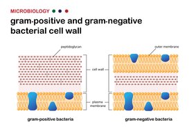
Cell Wall Structure and Function
The bacterial cell wall is essential for maintaining cell shape, protecting against osmotic stress, and is a major target for antibiotics.
Glycocalyx: Outer layer; capsule (organized, protective) vs. slime layer (loose, unstructured).
Flagella: Motility structures; composed of filament, hook, and basal body.
Axial Filaments: Endoflagella in spirochetes, enabling corkscrew movement.
Fimbriae and Pili: Attachment and genetic exchange structures.
Peptidoglycan: Polymer of N-acetylglucosamine (NAG) and N-acetylmuramic acid (NAM).
Gram-Positive vs. Gram-Negative Cell Walls: Gram-positive have thick peptidoglycan; Gram-negative have thin peptidoglycan and an outer membrane containing lipopolysaccharide (LPS).
LPS, Lipid A, O Polysaccharide: Components of Gram-negative outer membrane; Lipid A is an endotoxin.
Mycolic Acid: Waxy substance in mycobacterial cell walls.
Example: Gram-negative bacteria are more resistant to antibiotics due to their outer membrane.
Membrane Transport and Osmotic Balance
Cells regulate the movement of substances across the plasma membrane to maintain homeostasis.
Fluid Mosaic Model: Describes the dynamic nature of the plasma membrane.
Passive vs. Active Transport: Passive transport (diffusion, osmosis) does not require energy; active transport does.
Isotonic, Hypotonic, Hypertonic Solutions: Affect cell volume and integrity.
Additional info: In a hypertonic solution, bacterial cells lose water and shrink (plasmolysis); in a hypotonic solution, they may burst (lysis).
Endospores and Organelles
Some bacteria form endospores for survival; eukaryotic cells contain specialized organelles.
Endospores: Highly resistant structures formed during sporulation; germinate under favorable conditions.
Organelles: Nucleus, endoplasmic reticulum (ER), Golgi apparatus, mitochondria, chloroplasts—each with specific functions.
Endosymbiotic Theory: Mitochondria and chloroplasts originated from symbiotic bacteria.
Additional info: Mitochondria and chloroplasts contain their own DNA and ribosomes, supporting the endosymbiotic theory.
