 Back
Microbiology Study Guide: Microscopy, Cell Domains, Staining, and Viruses
Study Guide - Smart Notes
 Tailored notes based on your materials, expanded with key definitions, examples, and context.
Q1. What are the main parts of a compound microscope and how do they contribute to magnification and focusing?
Background
Topic: Microscopy
This question tests your understanding of the structure and function of a compound microscope, including how magnification is achieved and how focusing works.
Key Terms and Formulas
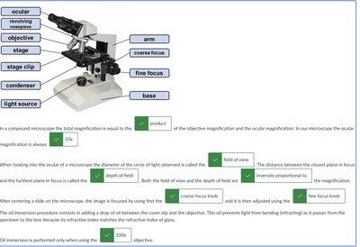
Ocular (eyepiece): The lens you look through, usually 10x magnification.
Objective lenses: Multiple lenses with different magnifications (e.g., 4x, 10x, 40x, 100x).
Stage: Platform where the slide is placed.
Coarse focus knob: Used for large adjustments in focus.
Fine focus knob: Used for small, precise adjustments.
Magnification formula:
Step-by-Step Guidance
Identify the main parts of the microscope: ocular, objectives, stage, condenser, light source, coarse and fine focus knobs.
Understand that total magnification is calculated by multiplying the magnification of the objective lens by the ocular lens.
When starting to focus, always use the lowest power objective (scanning power, usually 4x) and the coarse focus knob first.
Once the specimen is in focus at low power, switch to higher objectives and use the fine focus knob for precise focusing.
Consider how increasing magnification affects the field of view and depth of field.
Try solving on your own before revealing the answer!
Final Answer:
The main parts of a compound microscope include the ocular, objective lenses, stage, condenser, light source, coarse focus, and fine focus. Total magnification is the product of the objective and ocular magnifications. Start with the scanning power and coarse focus, then move to higher objectives and fine focus for detailed viewing.
Q2. How do field of view and depth of field change as magnification increases in microscopy?
Background
Topic: Microscopy - Field of View and Depth of Field
This question tests your understanding of how increasing magnification affects what you see through the microscope, specifically the diameter of the visible area (field of view) and the thickness of the specimen that remains in focus (depth of field).
Key Terms and Formulas
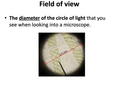
Field of View: The diameter of the circle of light you see when looking into the microscope.
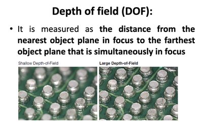
Depth of Field: The distance from the nearest object plane in focus to the farthest object plane that is simultaneously in focus.
Relationship: (inversely proportional)
Step-by-Step Guidance
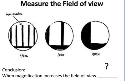
Recall that as you increase the magnification, the field of view decreases. This means you see a smaller area of the specimen.
Understand that depth of field also decreases with higher magnification, so only a thinner layer of the specimen remains in focus.
Visualize this by comparing what you see at 40x, 100x, and 400x magnification.
Think about why these changes occur: higher magnification zooms in on a smaller area and focuses on a thinner slice.
Try solving on your own before revealing the answer!
Final Answer:
As magnification increases, both the field of view and depth of field decrease. You see a smaller area and a thinner section of the specimen in focus. This is because higher magnification narrows the visible and focused region.
Q3. What are the three domains of life and how are prokaryotes and eukaryotes distinguished?
Background
Topic: Microbial Classification
This question tests your understanding of the classification of life into domains and the differences between prokaryotic and eukaryotic cells.
Key Terms and Formulas
Bacteria: Prokaryotic domain, no true nucleus.
Archaea: Prokaryotic domain, no true nucleus, distinct from bacteria.
Eukarya: Eukaryotic domain, cells have a true nucleus.
Prokaryotes: Cells without a true nucleus (Bacteria and Archaea).
Eukaryotes: Cells with a true nucleus (Eukarya).
Step-by-Step Guidance
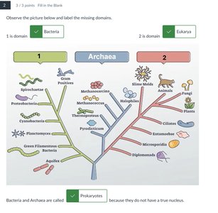
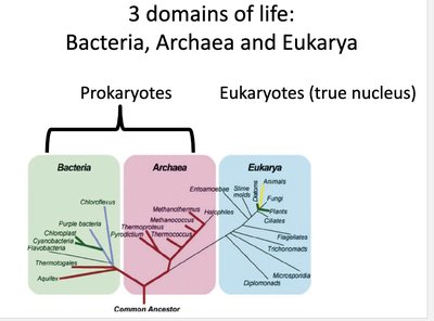
Identify the three domains: Bacteria, Archaea, and Eukarya.
Recognize that Bacteria and Archaea are prokaryotes, meaning they lack a true nucleus.
Eukarya are eukaryotes, meaning their cells contain a true nucleus.
Understand the evolutionary relationships and differences in cell structure between these domains.
Try solving on your own before revealing the answer!
Final Answer:
The three domains of life are Bacteria, Archaea, and Eukarya. Bacteria and Archaea are prokaryotes (no true nucleus), while Eukarya are eukaryotes (cells with a true nucleus).
Q4. What is oil immersion and why is it used at high magnification in microscopy?
Background
Topic: Microscopy - Oil Immersion
This question tests your understanding of the purpose and function of oil immersion in microscopy, especially at high magnifications (1000x).
Key Terms and Formulas
Oil Immersion: Technique used with the 100x objective lens to improve resolution.
Refractive Index: Measure of how much light bends when passing through a medium.
Resolution: Ability to distinguish two points as separate.
Step-by-Step Guidance
Recall that air and glass have different refractive indices, causing light to bend and scatter, which reduces resolution at high magnification.
Oil immersion uses a special oil with the same refractive index as glass, minimizing light bending and loss.
This technique is used only with the 100x objective lens, resulting in a total magnification of 1000x.
Understand that oil immersion improves image clarity and resolution by allowing more light to enter the objective lens.

Try solving on your own before revealing the answer!
Final Answer:
Oil immersion is used at 1000x magnification to improve resolution by reducing light loss and bending. The oil matches the refractive index of glass, resulting in clearer, sharper images at high magnification.
Q5. What are the main bacterial cell shapes and arrangements?
Background
Topic: Bacterial Morphology
This question tests your knowledge of the different shapes and arrangements of bacterial cells, which are important for identification and classification.
Key Terms and Formulas
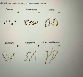
Coccus: Spherical shape.
Bacillus: Rod-shaped.
Vibrio: Curved rod.
Spirillum: Spiral-shaped.
Spirochete: Flexible spiral.
Branching filaments: Filamentous shape.
Arrangements: Single, pairs (diplo), chains (strepto), clusters (staphylo).
Step-by-Step Guidance
Identify the basic shapes: coccus, bacillus, vibrio, spirillum, spirochete, branching filaments.
Recognize common arrangements: single cells, pairs, chains, clusters.
Understand how these shapes and arrangements help in bacterial identification.
Review visual examples to reinforce recognition of each type.

Try solving on your own before revealing the answer!
Final Answer:
Main bacterial shapes include coccus, bacillus, vibrio, spirillum, spirochete, and branching filaments. Arrangements include singles, pairs, chains, and clusters, which are key for identification.
Q6. What is simple staining and how does it differ from positive and negative staining?
Background
Topic: Bacterial Staining Techniques
This question tests your understanding of simple staining methods and the difference between positive and negative stains.
Key Terms and Formulas
Simple Staining: Uses one dye to color cells.
Positive Stain: Basic dye stains cells darker than the background.
Negative Stain: Acidic dye stains the background, leaving cells lighter.
Common dyes: Safranin, methylene blue.
Step-by-Step Guidance
Understand that simple staining uses a single dye to add color to cells for better visibility.
Positive stains use basic dyes that bind to negatively charged cell surfaces, making cells appear darker.
Negative stains use acidic dyes that are repelled by cell surfaces, staining the background instead.
Review examples of each staining method and their appearance under the microscope.


Try solving on your own before revealing the answer!
Final Answer:
Simple staining uses one dye. Positive stains color cells darker than the background, while negative stains color the background, leaving cells lighter. This helps visualize cell shape and arrangement.
Q7. What are the steps and purpose of Gram staining?
Background
Topic: Gram Staining - Differential Staining
This question tests your knowledge of the Gram staining procedure, its steps, and how it differentiates between Gram-positive and Gram-negative bacteria.
Key Terms and Formulas
Gram Stain: Complex, differential staining method.
Primary stain: Crystal violet.
Mordant: Gram's iodine.
Decolorizer: Alcohol or acetone.
Counterstain: Safranin.
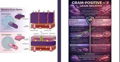
Gram-positive: Retains crystal violet, appears purple.
Gram-negative: Loses crystal violet, takes up safranin, appears pink/red.
Step-by-Step Guidance
Prepare a bacterial smear and heat fix it.
Apply crystal violet (primary stain) for one minute, then rinse with water.
Apply Gram's iodine (mordant) for one minute, then rinse with water.
Apply decolorizer (alcohol or acetone) briefly, then rinse with water.
Apply safranin (counterstain) for one minute, then rinse and observe.


Try solving on your own before revealing the answer!
Final Answer:
Gram staining involves applying crystal violet, iodine, decolorizer, and safranin in sequence. Gram-positive cells retain the purple dye, while Gram-negative cells take up the pink/red counterstain. This method distinguishes bacteria based on cell wall structure.
Q8. What is endospore staining and which bacteria form endospores?
Background
Topic: Endospore Staining
This question tests your understanding of the endospore staining technique and the types of bacteria that produce endospores.
Key Terms and Formulas
Endospore: A resistant, dormant structure formed by some bacteria.
Primary stain: Malachite green.
Counterstain: Safranin.
Bacillus and Clostridium: Genera that form endospores.
Diseases: Anthrax, tetanus, botulism, gas gangrene.
Step-by-Step Guidance
Prepare a bacterial smear and heat fix it.
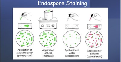
Apply malachite green and heat to drive the stain into endospores.
Rinse with water to remove stain from vegetative cells.
Apply safranin to stain vegetative cells pink/red.
Identify which bacteria form endospores and the diseases they cause.
Try solving on your own before revealing the answer!
Final Answer:
Endospore staining uses malachite green and heat, followed by safranin. Bacillus and Clostridium genera form endospores, causing diseases like anthrax, tetanus, botulism, and gas gangrene.
Q9. What are selective and differential media, and how do Mannitol and MacConkey agars work?
Background
Topic: Microbial Growth Media
This question tests your understanding of selective and differential media, specifically Mannitol Salt Agar (MSA) and MacConkey Agar.
Key Terms and Formulas
Selective Media: Allows growth of certain microbes while inhibiting others.
Differential Media: Distinguishes between microbes based on biochemical reactions.
MSA: Selects for halophilic bacteria, differentiates based on mannitol fermentation.
MacConkey: Selects for Gram-negative bacteria, differentiates based on lactose fermentation.
Step-by-Step Guidance
Understand that MSA contains high salt, selecting for salt-loving (halophilic) bacteria.
MSA differentiates based on mannitol fermentation; acid production turns the pH indicator yellow.
MacConkey contains crystal violet and bile salts, selecting for Gram-negative bacteria.
MacConkey differentiates based on lactose fermentation; acid production turns the pH indicator pink.

Try solving on your own before revealing the answer!
Final Answer:
MSA selects for halophilic bacteria and differentiates based on mannitol fermentation (yellow color). MacConkey selects for Gram-negative bacteria and differentiates based on lactose fermentation (pink color).
Q10. How are viruses classified as enveloped or naked, and what are their genome types?
Background
Topic: Virology - Virus Structure and Classification
This question tests your understanding of virus classification based on envelope presence and genome type.
Key Terms and Formulas
Enveloped Virus: Has a lipid membrane surrounding the capsid.
Naked Virus: Lacks a lipid envelope.
Genome Types: ssRNA (single-stranded RNA), dsDNA (double-stranded DNA), segmented, linear, circular.
Step-by-Step Guidance
Identify which viruses are enveloped and which are naked based on their structure.
Classify viruses by their genome type: negative or positive sense RNA, segmented or non-segmented, linear or circular DNA.
Understand how these features affect virus infectivity and classification.
Try solving on your own before revealing the answer!
Final Answer:
Viruses are classified as enveloped or naked based on the presence of a lipid envelope. Genome types include negative or positive single-stranded RNA (segmented or non-segmented), and double-stranded DNA (linear or circular).
