 Back
BackMicrobiology Study Guide: Prokaryotes, Eukaryotes, and Viruses
Study Guide - Smart Notes
 Tailored notes based on your materials, expanded with key definitions, examples, and context.
General Characteristics of Prokaryotic Organisms
Prokaryotic Cell Structure and Diversity
Prokaryotes are unicellular organisms lacking a nucleus and membrane-bound organelles. They are the most numerous and diverse cellular microbes, thriving in various habitats with sufficient moisture. Only a few prokaryotes cause disease in humans, and archaea are not known to cause disease. The endosymbiotic theory suggests mitochondria are derived from prokaryotes, as evidenced by the similarity of 70S ribosomes in prokaryotes and mitochondria.
Ribosomes: Sites of protein synthesis; prokaryotes have 70S ribosomes, eukaryotes have 80S.
Treatments: More available for prokaryotic diseases due to lower toxicity compared to eukaryotic diseases.
Archaea: Not known to cause disease.
Reproduction of Prokaryotic Cells
All prokaryotes reproduce asexually, using several methods:
Binary fission: Most common; one cell divides into two.
Snapping division: Variation of binary fission.
Reproductive spores: Similar to fungi; source of antibiotics.
Budding: New cell grows from parent.
Viviparity: Live offspring emerge from the mother cell (e.g., Epulopiscium).
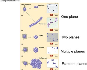
Arrangements of Prokaryotic Cells
The arrangement of prokaryotic cells results from the planes in which cells divide and whether daughter cells separate. Microscopy is used to group bacteria, aiding diagnosis.
Bacilli: Most abundant shape; often appear single or in chains (streptobacilli).
Cocci: Many arrangements based on division planes:
One plane: Diplococci (pairs), Streptococci (chains)
Two planes: Tetrads (squares)
Multiple planes: Sarcinae (cubical packets)
Random planes: Staphylococci (clusters)
Modern Prokaryotic Classification
Classification is based on genetic relatedness of rRNA sequences, dividing life into three domains: Archaea, Bacteria, and Eukarya. Bergey’s Manual provides a systematic classification scheme.
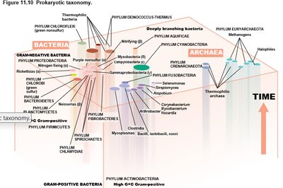
Survey of Bacteria and Archaea
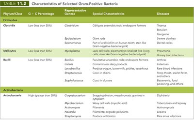
Low G + C Gram-Positive Bacteria
These bacteria have less than 50% guanine-cytosine content and are classified in the phylum Firmicutes. They include medically important genera such as Clostridia, Mycoplasmas, Bacillus, Listeria, Lactobacillus, Streptococcus, Enterococcus, and Staphylococcus.
Clostridia: Spore-forming, obligate anaerobes; cause tetanus, botulism, gas gangrene, and C. diff infections.
Mycoplasmas: Lack cell walls; cause pneumonia and urinary tract infections; "fried egg" appearance in culture.
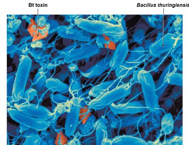
Bacillus: Endospore-forming aerobes; includes Bacillus thuringiensis (insecticide) and Bacillus anthracis (anthrax).
Listeria: Psychrophile; contaminates refrigerated foods; can cause miscarriage in pregnant women.
Lactobacillus: Normal flora; used in yogurt and probiotics.
Streptococcus/Enterococcus: Cause cavities, pneumonia, and opportunistic infections.
Staphylococcus: Common skin flora; produces toxins and enzymes; causes wound and heart infections.
Phylum/Class | G + C Percentage | Representative Genera | Special Characteristics | Diseases |
|---|---|---|---|---|
Firmicutes: Clostridia | Low (<50%) | Clostridium | Obligate anaerobic rod, endospore formers | Tetanus, botulism, gas gangrene, severe diarrhea |
Firmicutes: Mollicutes | Low (<50%) | Mycoplasma | Lack cell wall, pleomorphic, smallest free-living cells | Pneumonia, urinary tract infections |
Firmicutes: Bacilli | Low (<50%) | Bacillus, Listeria, Lactobacillus, Streptococcus, Staphylococcus | Endospore formers, produce yogurt, butter, pickles, asexual spores, cocci in clusters | Anthrax, food poisoning, wound infections, pneumonia, strep throat, sinus infections |
Actinobacteria | High (>50%) | Corynebacterium, Mycobacterium, Actinomyces, Nocardia, Streptomyces | Pleomorphic, filaments, degrade pollutants, produce antibiotics | Diphtheria, tuberculosis, leprosy, rare sinus infections |
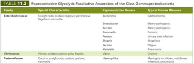
Gram-Negative Bacteria: Proteobacteria
Proteobacteria are the largest and most diverse group of Gram-negative bacteria, divided into five classes: Alpha-, Beta-, Gamma-, Delta-, and Epsilonproteobacteria.
Alphaproteobacteria: Includes Rickettsia (typhus), Brucella (brucellosis), and Agrobacterium (plant tumors).
Betaproteobacteria: Includes Neisseria (meningitis, gonorrhea), Bordetella (whooping cough), Burkholderia (lung infections).
Gammaproteobacteria: Includes Legionella (Legionnaire’s disease), Coxiella (Q fever), Enterobacteriaceae (GI tract), Vibrioceae (cholera), Pasteurellaceae (Haemophilus).
Pseudomonads: Soil bacteria; cause nosocomial infections; resistant to antibiotics.
Epsilonproteobacteria: Includes Campylobacter (blood poisoning), Helicobacter pylori (ulcers, gastric cancer).
Family | Special Characteristics | Representative Genera | Typical Human Diseases |
|---|---|---|---|
Enterobacteriaceae | Straight rods, oxidase negative, peritrichous flagella or nonmotile | Escherichia, Enterobacter, Serratia, Salmonella, Proteus, Shigella, Yersinia | Rarely pathogenic, gastroenteritis, urinary tract infection, plague, shigellosis |
Vibrionaceae | Vibrios, oxidase positive, polar flagella | Vibrio | Cholera |
Pasteurellaceae | Cocci or straight rods, oxidase positive, nonmotile | Haemophilus | Meningitis in children, middle ear infections, pneumonia |
Characterizing and Classifying Eukaryotes
General Characteristics of Eukaryotic Microorganisms
Eukaryotic microorganisms include protozoa, fungi, algae, and water molds. They are more complex than prokaryotes, with DNA packaged as chromosomes in a nucleus. Eukaryotes reproduce both asexually and sexually, forming gametes and zygotes.
Reproduction of Eukaryotes
Meiosis: Reduces chromosome number by half.
Mitosis: Produces identical daughter cells.
Cytokinesis: Division of cytoplasm; may be delayed in coenocytic cells (multinucleated).
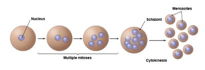
Schizogony: Multiple mitoses followed by cytokinesis, producing merozoites (seen in protozoa).
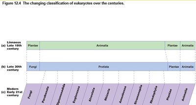
Classification of Eukaryotes
Classification has evolved from two kingdoms (Plantae and Animalia) to include Fungi, Protista, and multiple modern groups based on rRNA sequences.
Protozoa
General Characteristics and Morphology
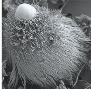
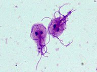
Protozoa are unicellular, eukaryotic organisms lacking a cell wall. They inhabit aquatic environments and are highly diverse in morphology and life cycle stages.
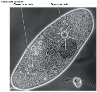
Contractile vacuoles: Pump water out to prevent cell rupture in hypotonic environments.
Motility: Move by cilia, flagella, or pseudopods; apicomplexans are non-motile.
Life cycle stages: Trophozoite (active, feeding), cyst (resting, dormant).
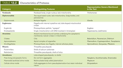
Classification of Protozoa
Protozoa are classified into six groups based on rRNA and distinguishing features.
Category | Distinguishing Features | Representative Genera |
|---|---|---|
Parabasala | Parabasal body, single nucleus, lack mitochondria | Trichomonas |
Diplomonadida | Two equal-sized nuclei, lack mitochondria, Golgi bodies, and peroxisomes | Giardia |
Euglenozoa | Flagella with internal crystalline rod, disk-shaped mitochondria | Euglena, Trypanosoma, Leishmania |
Alveolates | Alveoli, cilia, apical complex of organelles | Balantidium, Plasmodium, Paramecium, Dinoflagellates |
Rhizaria | Threadlike pseudopodia, shells of silica | Foraminifera, Radiolaria |
Amoebozoa | Lobe-shaped pseudopodia | Naegleria, Acanthamoeba, Entamoeba |
Fungi
General Characteristics and Significance
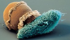
Fungi are chemoheterotrophic eukaryotes with cell walls composed of chitin. They decompose dead organisms, recycle nutrients, and are used in food and drug production. About 30% cause diseases (mycoses) in plants, animals, and humans.
Morphology of Fungi
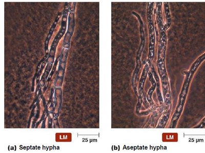
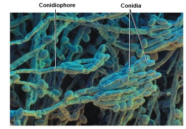
Hyphae: Filamentous structures; may be septate (divided) or aseptate (coenocytic).
Mycelium: Mass of hyphae.
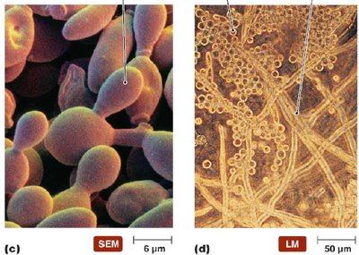
Dimorphism: Ability to change shape based on temperature; yeast at 37°C, mold at room temperature.
Pseudohyphae: Yeast cells that fail to separate completely.
Reproduction of Fungi
Asexual reproduction: Mitosis and cytokinesis; budding in yeasts, fragmentation in molds.
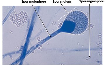
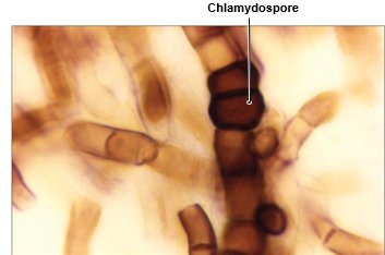
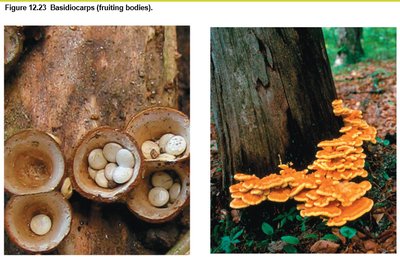
Spore formation: Asexual spores (sporangiospores, conidia, chlamydospores); sexual spores (zygospores, ascospores, basidiospores).
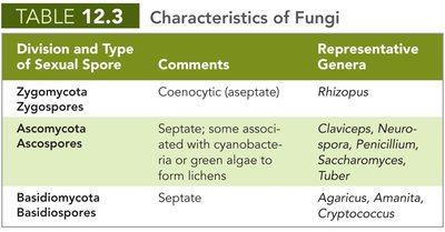
Classification of Fungi
Division and Type of Sexual Spore | Comments | Representative Genera |
|---|---|---|
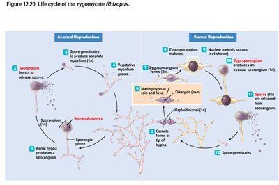
Zygomycota (Zygospores) | Coenocytic (aseptate) | Rhizopus |
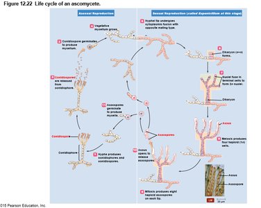
Ascomycota (Ascospores) | Septate; some associated with cyanobacteria or green algae to form lichens | Claviceps, Neurospora, Penicillium, Saccharomyces, Tuber |
Basidiomycota (Basidiospores) | Septate | Agaricus, Amanita, Cryptococcus |
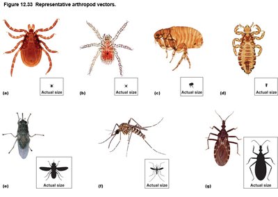
Other Eukaryotes: Parasitic Helminths and Arthropod Vectors
Arthropod Vectors
Arthropods such as ticks, fleas, lice, flies, mosquitoes, and kissing bugs serve as mechanical or biological vectors for various pathogens, including protozoa and viruses.
Characteristics of Viruses, Viroids, and Prions
Are Viruses Alive?
Viruses are miniscule, acellular infectious agents composed of protein and DNA or RNA. They cannot carry out metabolic pathways, grow, or reproduce independently, and must recruit host cell machinery for replication.
Virion: Extracellular state; protein coat (capsid) surrounds nucleic acid.
Capsid: Composed of capsomeres; provides protection and attachment to host cells.
Envelope: Acquired from host cell membrane; contains viral glycoproteins (spikes).
Genetic material: DNA or RNA, never both; may be single- or double-stranded, linear or circular.
Classification and Replication of Viruses
Classification: Based on nucleic acid, envelope presence, shape, and size.
Lytic cycle: Attachment, entry, synthesis, assembly, release; results in host cell lysis.
Lysogeny: Viral genome integrates into host DNA; host reproduces normally until lysis.
Animal viruses: Entry via direct penetration, membrane fusion, or endocytosis; DNA viruses replicate in nucleus, RNA viruses in cytoplasm.
Latency: Animal viruses may remain dormant in host cells for years.
Viroids and Prions
Viroids: Infectious RNA molecules lacking capsid; infect plants.
Prions: Proteinaceous infectious particles; cause spongiform encephalopathies in mammals.
Infection, Infectious Diseases, and Epidemiology
Symbiotic Relationships
Mutualism: Both organisms benefit (e.g., E. coli in human colon).
Commensalism: One benefits, other unaffected (e.g., Staphylococcus epidermidis on skin).
Parasitism: One benefits, other harmed (e.g., Mycobacterium tuberculosis in human lung).
Normal Microbiota
Resident microbiota: Present throughout life; mostly commensal.
Transient microbiota: Temporary; outcompeted by resident flora.
Opportunistic pathogens: Cause disease when introduced to unusual sites or in immunocompromised hosts.
Reservoirs of Infection
Animal reservoirs: Zoonoses; diseases spread from animals to humans.
Human carriers: Asymptomatic individuals can transmit disease.
Nonliving reservoirs: Soil, water, food.
Portals of Entry and Exit
Entry: Skin, mucous membranes, placenta, parenteral route.
Exit: Same as entry; pathogens leave in secretions or excretions.
Manifestations and Etiology of Disease
Symptoms: Subjective (e.g., nausea).
Signs: Objective (e.g., vomiting).
Syndrome: Group of symptoms/signs.
Koch’s Postulates: Criteria for identifying causative agents of disease.
Virulence Factors
Adhesion: Attachment to host cells via fimbriae, spikes, or biofilms.
Biofilms: Bacterial communities communicating via quorum sensing.
Extracellular enzymes: Break down host barriers (e.g., hyaluronidase, collagenase).
Toxins: Endotoxins (LPS), exotoxins (proteins).
Antiphagocytic factors: Capsules, chemicals preventing phagocytosis.
Stages of Infectious Disease
Incubation period
Prodromal period
Illness
Decline
Convalescence
Modes of Transmission
Contact transmission: Direct, indirect, droplet.
Vehicle transmission: Airborne, waterborne, foodborne, bodily fluids.
Vector transmission: Biological (mosquitoes, ticks), mechanical (flies).
Epidemiology
Incidence: Number of new cases.
Prevalence: Total cases (new and existing).
Endemic: Stable incidence.
Sporadic: Few cases.
Epidemic: Higher frequency than usual.
Pandemic: Disease crosses continents.
Additional info: Academic context and explanations were added to ensure completeness and clarity for exam preparation.
