 Back
BackMicrobiology Study Guide: Spirochetes and Gram-Negative Bacteria
Study Guide - Smart Notes
 Tailored notes based on your materials, expanded with key definitions, examples, and context.
Q1. What are the stages of syphilis and their associated clinical signs?
Background
Topic: Spirochetes – Treponema pallidum (Syphilis)
This question tests your understanding of the progression of syphilis, a disease caused by the spirochete Treponema pallidum. You should be able to identify the clinical signs associated with each stage and understand how the disease is diagnosed and treated.
Key Terms:
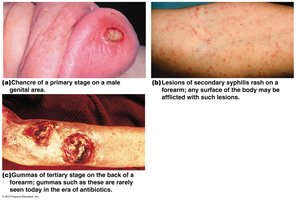
Primary stage: Characterized by a chancre (painless ulcer).
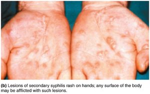
Secondary stage: Rash, often on palms and soles, and other systemic symptoms.
Latent stage: No visible symptoms.
Tertiary stage: Gummas, neurosyphilis, cardiovascular complications.
Step-by-Step Guidance
Review the morphology and transmission routes of Treponema pallidum. Remember it is a spirochete transmitted sexually or congenitally.
Identify the clinical sign of the primary stage: Look for the presence of a chancre at the site of infection.
Describe the secondary stage: Note the appearance of a rash, which can occur on various surfaces of the body, including the palms and soles.
Understand the latent stage: Recognize that this stage is asymptomatic, but the infection persists.
Consider the tertiary stage: Identify gummas and neurological symptoms, which are rare today due to antibiotic treatment.
Try solving on your own before revealing the answer!
Final Answer:
Primary stage: Chancre; Secondary stage: Rash; Latent stage: No symptoms; Tertiary stage: Gummas/neurosyphilis.
Each stage has distinct clinical features, and diagnosis is confirmed by serological testing for antibodies against T. pallidum.
Q2. What is Hemolytic Uremic Syndrome (HUS) and how is E. coli O157:H7 involved?
Background
Topic: Gram-Negative Bacteria – E. coli O157:H7
This question tests your knowledge of overt pathogens, specifically E. coli O157:H7, and its role in causing HUS, a serious complication involving hemolytic anemia and kidney damage.
Key Terms:
Hemolytic Uremic Syndrome (HUS): A condition characterized by hemolytic anemia, acute kidney injury, and thrombocytopenia.
E. coli O157:H7: A strain of E. coli that produces Shiga-toxin, leading to HUS.
Transmission: Fecal-oral route, often through contaminated food (e.g., beef) or petting zoos.
Step-by-Step Guidance
Recall the normal habitat of E. coli O157:H7 (cows) and its transmission routes.
Understand the role of Shiga-toxin in damaging red blood cells and causing kidney injury.
Identify the clinical signs of HUS: hemolytic anemia, kidney failure, and colitis.
Consider the importance of laboratory diagnosis: stool culture and serotyping for E. coli O157:H7.
Try solving on your own before revealing the answer!
Final Answer:
HUS is caused by E. coli O157:H7 through Shiga-toxin production, leading to hemolytic anemia and kidney damage. Transmission is via contaminated food or contact with animals.
Diagnosis is confirmed by stool culture and serotyping.
Q3. What are the clinical manifestations and laboratory diagnosis of Pseudomonas aeruginosa infections?
Background
Topic: Gram-Negative Bacilli – Pseudomonas aeruginosa
This question tests your understanding of opportunistic infections caused by Pseudomonas aeruginosa, especially in hospital settings and burn patients.
Key Terms:
Pseudomonas aeruginosa: An environmental, opportunistic pathogen.
Clinical manifestations: UTI, post-burn wound infections, dermatitis, swimmer’s ear.
Lab diagnosis: Gram stain, culture, biochemical tests; blue-green pigment on TSA.
Step-by-Step Guidance
Identify the types of infections caused by P. aeruginosa, especially in immunocompromised or hospitalized patients.
Describe the characteristic appearance of infected wounds (e.g., post-burn) and the blue-green pigment produced by the bacterium.
Review laboratory diagnostic methods: Gram stain, culture on TSA, and biochemical tests.
Consider antibiotic resistance as a challenge in treatment.

Try solving on your own before revealing the answer!
Final Answer:
Pseudomonas aeruginosa causes post-burn infections, UTIs, and other opportunistic infections. Diagnosis involves Gram stain, culture, and detection of blue-green pigment. Treatment is complicated by antibiotic resistance.
Q4. How does Helicobacter pylori cause peptic ulcers and how is it diagnosed?
Background
Topic: Curved Gram-Negative Rods – Helicobacter pylori
This question tests your understanding of the pathogenesis of peptic ulcers and stomach cancer caused by H. pylori, as well as laboratory diagnostic methods.
Key Terms:
Helicobacter pylori: Curved, gram-negative rod.
Urease enzyme: Breaks down urea into ammonia, neutralizing stomach acid.
Diagnosis: Serology for antigens/antibodies, stool antigen test, gastric biopsy.
Step-by-Step Guidance
Describe how H. pylori colonizes the stomach lining and survives acidic conditions by producing urease.
Explain how the ammonia produced by urease activity damages the mucosal layer, leading to ulcers.
Review laboratory diagnostic methods: serology, stool antigen test, and gastric biopsy.
Try solving on your own before revealing the answer!
Final Answer:
H. pylori causes peptic ulcers by producing urease, which neutralizes stomach acid and damages the mucosa. Diagnosis is made by serology, stool antigen test, or gastric biopsy.
Q5. What is the life cycle of Vibrio cholerae and how does it cause disease?
Background
Topic: Curved Gram-Negative Rods – Vibrio cholerae
This question tests your understanding of the transmission, pathogenesis, and clinical features of cholera, a disease caused by V. cholerae.
Key Terms:
Vibrio cholerae: Curved, gram-negative rod.
Cholera exotoxin: Causes massive watery diarrhea (rice-water stools).
Transmission: Fecal-oral route, contaminated water or food.
Step-by-Step Guidance
Describe the environmental reservoirs and transmission routes of V. cholerae.
Explain how the bacterium adheres to and penetrates the mucous layer of the intestine.
Understand the mechanism of cholera toxin in causing watery diarrhea.
Review the importance of fluid and electrolyte replacement in treatment.

Try solving on your own before revealing the answer!
Final Answer:
V. cholerae is transmitted via contaminated water/food, adheres to the intestinal mucosa, and produces cholera toxin, resulting in rice-water diarrhea. Treatment includes antibiotics and fluid/electrolyte replacement.
Q6. How is Neisseria gonorrhoeae identified in the laboratory and what diseases does it cause?
Background
Topic: Gram-Negative Diplococcus – Neisseria gonorrhoeae
This question tests your knowledge of the laboratory identification and clinical manifestations of gonorrhea caused by N. gonorrhoeae.
Key Terms:
Neisseria gonorrhoeae: Gram-negative diplococcus.
Diseases: Gonorrhea, cervicitis, pelvic inflammatory disease, ophthalmic neonatorum, arthritis.
Lab diagnosis: Gram stain showing diplococci inside neutrophils.
Step-by-Step Guidance
List the diseases caused by N. gonorrhoeae.
Describe the laboratory identification: Gram stain of urethral exudate, look for gram-negative diplococci inside neutrophils.
Understand the importance of rapid diagnosis for effective treatment.
Try solving on your own before revealing the answer!
Final Answer:
N. gonorrhoeae is identified by Gram stain showing diplococci inside neutrophils and causes gonorrhea and related diseases.
Q7. What are the warning signs of meningococcal disease caused by Neisseria meningitidis?
Background
Topic: Gram-Negative Diplococcus – Neisseria meningitidis
This question tests your ability to recognize the clinical signs and laboratory diagnosis of meningococcal disease.
Key Terms:
Neisseria meningitidis: Gram-negative diplococcus.
Meningococcal disease: Meningitis, sepsis.
Warning signs: Severe headache, fever, stiff neck, rash, rapid breathing, drowsiness.
Lab diagnosis: Gram stain, capsule antigen testing, culture of spinal fluid or blood.
Step-by-Step Guidance
List the classic and early warning signs of meningococcal disease.
Describe the laboratory diagnostic methods: Gram stain, capsule antigen testing, culture.
Understand the importance of rapid diagnosis and treatment.

Try solving on your own before revealing the answer!
Final Answer:
Warning signs include severe headache, fever, stiff neck, rash, and rapid breathing. Diagnosis is made by Gram stain, capsule antigen testing, and culture.
