 Back
BackMicrobiology Study Guide: Staining Techniques and Microbial Morphology
Study Guide - Smart Notes
 Tailored notes based on your materials, expanded with key definitions, examples, and context.
Gram Staining
Introduction to Gram Staining
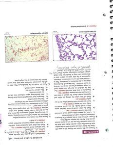
Gram staining is a fundamental technique in microbiology used to differentiate bacterial species into two groups: Gram-positive and Gram-negative. This distinction is based on differences in cell wall structure and is crucial for bacterial identification and classification.
Gram-positive bacteria: Retain the crystal violet stain and appear purple due to a thick peptidoglycan layer.
Gram-negative bacteria: Lose the crystal violet stain and take up the counterstain (safranin), appearing pink/red due to a thinner peptidoglycan layer and an outer membrane.
Applications: Used in clinical diagnostics, research, and guiding antibiotic therapy.
Gram Staining Procedure
The Gram stain involves a series of steps using specific reagents. Each step is essential for accurate differentiation.
Prepare a heat-fixed smear of the bacterial sample.
Apply crystal violet (primary stain) for 1 minute.
Rinse with water.
Add iodine (mordant) for 1 minute.
Rinse with water.
Decolorize with alcohol for 10-20 seconds.
Rinse with water.
Counterstain with safranin for 1 minute.
Rinse with water and blot dry.
Example: Staphylococcus aureus (Gram-positive) and Escherichia coli (Gram-negative) are commonly used in Gram stain demonstrations. 
Acid-Fast Staining
Introduction to Acid-Fast Staining
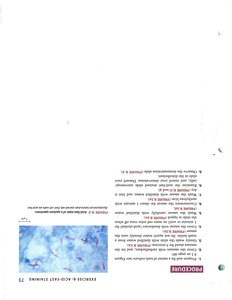
Acid-fast staining is used to identify bacteria with waxy cell walls, such as mycobacteria. These bacteria resist decolorization by acid-alcohol due to the presence of mycolic acids.
Acid-fast bacteria: Retain the primary stain (carbolfuchsin) and appear red.
Non-acid-fast bacteria: Lose the primary stain and take up the counterstain (methylene blue), appearing blue.
Applications: Essential for diagnosing tuberculosis and leprosy.
Acid-Fast Staining Procedure
Prepare a heat-fixed smear.
Apply carbolfuchsin for 5 minutes.
Rinse with water.
Decolorize with acid-alcohol for 1 minute.
Rinse with water.
Counterstain with methylene blue for 1 minute.
Rinse with water and blot dry.
Example: Mycobacterium tuberculosis is acid-fast, while Staphylococcus aureus is non-acid-fast. 
Structural Stains: Endospore, Capsule, and Flagella
Introduction to Structural Stains
Structural stains are specialized techniques used to visualize bacterial structures such as endospores, capsules, and flagella. These features are important for bacterial survival, pathogenicity, and motility.
Endospore stain: Detects dormant, resistant spores within bacteria.
Capsule stain: Reveals the presence of a protective polysaccharide layer.
Flagella stain: Highlights bacterial motility structures.
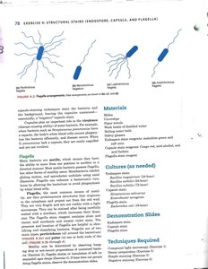
Flagella Arrangements
Bacteria can have different flagella arrangements, which are important for identification.
Monotrichous: Single flagellum at one end.
Lophotrichous: Cluster of flagella at one end.
Amphitrichous: Flagella at both ends.
Peritrichous: Flagella distributed over the entire cell surface.
Endospore Staining Procedure
Apply malachite green to heat-fixed smear and steam for 5 minutes.
Rinse with water.
Counterstain with safranin for 1 minute.
Rinse and blot dry.
Example: Bacillus and Clostridium species produce endospores. 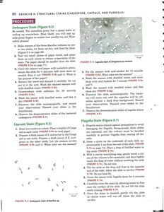
Capsule Staining Procedure
Mix bacteria with India ink or nigrosin on a slide.
Spread and air dry.
Stain with crystal violet for 1 minute.
Rinse and blot dry.
Example: Klebsiella pneumoniae produces a prominent capsule. 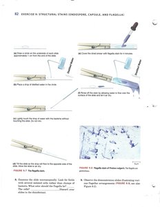
Flagella Staining Procedure
Apply flagella stain to heat-fixed smear for 4-5 minutes.
Rinse with water.
Blot dry and examine under oil immersion.
Example: Proteus vulgaris is highly motile with peritrichous flagella. 
Morphological Unknowns
Identifying Unknown Microbes by Morphology
Morphological analysis is a key step in identifying unknown bacteria. Staining techniques, cell shape, arrangement, and structural features are used to narrow down possible species.
Cell shapes: Cocci (spherical), bacilli (rod-shaped), spirilla (spiral-shaped).
Arrangements: Chains, clusters, pairs, etc.
Structural features: Presence of endospores, capsules, flagella.
Example: A Gram-positive cocci in clusters with a capsule may be Staphylococcus species.
Summary Table: Staining Techniques
Stain | Target Structure | Primary Dye | Counterstain | Result |
|---|---|---|---|---|
Gram Stain | Cell wall | Crystal violet | Safranin | Purple (Gram+), Pink (Gram-) |
Acid-Fast Stain | Mycolic acid cell wall | Carbolfuchsin | Methylene blue | Red (Acid-fast), Blue (Non-acid-fast) |
Endospore Stain | Endospores | Malachite green | Safranin | Green (spores), Red (cells) |
Capsule Stain | Capsule | India ink/Nigrosin | Crystal violet | Clear halo (capsule) |
Flagella Stain | Flagella | Flagella stain | None | Visible flagella |
Key Terms and Definitions
Staining: The process of adding dyes to microbial cells to enhance visibility under a microscope.
Mordant: A substance that helps fix the dye to the cell structure.
Decolorizer: Removes the primary stain from certain cells, allowing differentiation.
Counterstain: A secondary dye used to provide contrast.
Endospore: A dormant, resistant structure formed by some bacteria.
Capsule: A gelatinous layer surrounding some bacteria, aiding in evasion of host defenses.
Flagella: Whip-like appendages used for bacterial motility.
Additional info: Academic context was added to clarify procedures, applications, and examples for each staining technique.
