 Back
BackMicrobiology Study Guide: The Microbial World, Microscopy, and Cell Structure
Study Guide - Smart Notes
 Tailored notes based on your materials, expanded with key definitions, examples, and context.
Chapter 1: The Microbial World and You
Historical Figures in Microbiology
Microbiology has been shaped by the discoveries of several key scientists whose work laid the foundation for modern understanding of microorganisms.
Robert Hooke: First described cells, marking the beginning of cell theory.
Anton van Leeuwenhoek: Observed "animcules" (microorganisms) using simple microscopes.
Louis Pasteur: Demonstrated fermentation, developed pasteurization, and disproved spontaneous generation with his S-shaped flask experiment.
Robert Koch: Formulated the germ theory of disease and established Koch’s postulates for identifying causative agents of disease.
Key Terms and Concepts
Microbiome (Microbiota): The community of microbes living stably on or within the human body, contributing to health and disease prevention.
Normal Microbiota: Microorganisms acquired and maintained in healthy humans.
Transient Microbiota: Microorganisms that temporarily colonize the human body.
Scientific Nomenclature: The system of naming organisms using Genus and Specific Epithet (e.g., Escherichia coli).
Three Domains: Bacteria, Archaea, and Eukarya—distinguished by cellular characteristics.
Peptidoglycan: A structural molecule in bacterial cell walls.
Chitin and Cellulose: Structural polysaccharides in fungi and plants, respectively.
Binary Fission: Asexual reproduction in prokaryotes.
Spontaneous Generation vs. Biogenesis: The historical debate over whether life arises from nonliving matter (spontaneous generation) or from existing life (biogenesis).
Cell Theory: All living things are composed of cells.
Germ Theory of Disease: Microorganisms cause disease.
Koch’s Postulates: Criteria for establishing a causal relationship between a microbe and a disease.
Emerging Infectious Diseases (EID): Newly identified or increasing diseases.
Core Concepts
Microbes play essential roles in ecosystems, such as nutrient cycling and symbiosis.
Scientific names are written with the genus capitalized and the species lowercase, both italicized.
Major microorganism groups are distinguished by cellular structure: Archaea (no peptidoglycan), Bacteria (peptidoglycan), Fungi (chitin), Protists, Algae, Prokaryotes, and Eukaryotes.
The shift from spontaneous generation to biogenesis was pivotal in scientific understanding.
The Golden Age of Microbiology led to advances in disease prevention and treatment.
Chapter 3: Observing Microorganisms Through a Microscope
Microscopy Fundamentals
Microscopy is essential for observing microorganisms, with various techniques offering different levels of magnification and resolution.
Micrometer (µm) and Nanometer (nm): Units for measuring microorganisms.
Total Magnification: Product of the objective and ocular lens magnifications.
Resolution (Resolving Power): Ability to distinguish two points as separate; depends on wavelength and numerical aperture.
Refractive Index: Measure of how light bends as it passes through substances; immersion oil increases resolution by reducing refraction.
Types of Microscopy
Brightfield Microscopy: Standard light microscopy; best for stained specimens.
Darkfield Microscopy: Enhances contrast in unstained samples; useful for live organisms.
Phase-Contrast Microscopy: Visualizes internal structures in living cells.
Fluorescence Microscopy: Uses fluorescent dyes to visualize specific structures.
Confocal and Scanning Acoustic Microscopy: Advanced techniques for detailed imaging.
TEM vs. SEM: Transmission Electron Microscopy (TEM) provides internal details; Scanning Electron Microscopy (SEM) shows surface structures.
Staining Techniques
Fixing: Preserves and attaches specimens to slides.
Simple Stain: Uses one dye to highlight cells.
Differential Stain: Distinguishes cell types (e.g., Gram stain).
Gram Stain: Involves primary stain, mordant, decolorizer, and counterstain to differentiate Gram-positive and Gram-negative bacteria.
Acid-Fast Stain: Identifies mycobacteria.
Capsule, Endospore, and Flagella Stains: Specialized stains for specific structures.
Core Concepts
Shorter wavelengths yield higher resolution in microscopy.
Light passes through lenses and specimen in a compound microscope.
Light and electron microscopes differ in resolution and magnification; electron microscopes are superior for ultrastructural details.
Basic dyes bind to negatively charged bacterial cell walls; acidic dyes stain background.
Differential staining is crucial for pathogen identification in clinical settings.
Chapter 4: Functional Anatomy of Prokaryotic and Eukaryotic Cells
Cell Types and Morphology
Microbial cells are classified as prokaryotes or eukaryotes, each with distinct structural features.
Prokaryote: Cells lacking a nucleus and membrane-bound organelles (e.g., bacteria, archaea).
Eukaryote: Cells with a nucleus and organelles (e.g., fungi, protists, algae).
Monomorphic: Single shape.
Pleomorphic: Variable shapes.
Coccus, Bacillus, Spiral: Common bacterial shapes; spiral includes Vibrio, Spirillum, Spirochete.
Diplococci, Staphylococci, Streptococci: Arrangements of cocci.
Cell Structures and Functions
Glycocalyx: Protective layer; capsule (organized) or slime layer (unorganized).
Flagella: Motility structures; composed of filament, hook, and basal body.
Axial Filaments: Endoflagella in spirochetes.
Fimbriae and Pili: Attachment and conjugation structures.
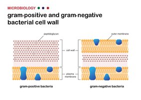
Peptidoglycan: Polymer of N-acetylglucosamine (NAG) and N-acetylmuramic acid (NAM); provides cell wall strength.
Gram-Positive vs. Gram-Negative Cell Walls: Gram-positive has thick peptidoglycan; Gram-negative has thin peptidoglycan and an outer membrane containing lipopolysaccharide (LPS).
LPS, Lipid A, O Polysaccharide: Components of Gram-negative cell wall; Lipid A is toxic.
Mycolic Acid: Waxy substance in mycobacterial cell walls.
Fluid Mosaic Model: Describes plasma membrane structure.
Passive vs. Active Transport: Movement of substances across membranes; passive does not require energy, active does.
Isotonic, Hypotonic, Hypertonic Solutions: Affect cell water balance.
Endospores: Resistant structures formed by some bacteria; sporulation and germination.
Organelles: Nucleus, endoplasmic reticulum (ER), Golgi apparatus, mitochondria, chloroplasts.
Endosymbiotic Theory: Mitochondria and chloroplasts originated from prokaryotic cells engulfed by ancestors of eukaryotes.
Comparison of Gram-Positive and Gram-Negative Cell Walls
The cell wall structure is a key distinguishing feature between Gram-positive and Gram-negative bacteria, affecting their staining properties, susceptibility to antibiotics, and pathogenicity.
Gram-Positive: Thick peptidoglycan layer, no outer membrane, teichoic acids present.
Gram-Negative: Thin peptidoglycan layer, outer membrane with LPS, periplasmic space.
Feature | Gram-Positive | Gram-Negative |
|---|---|---|
Peptidoglycan Thickness | Thick | Thin |
Outer Membrane | Absent | Present |
LPS (Lipopolysaccharide) | Absent | Present |
Teichoic Acids | Present | Absent |
Stain Color (Gram Stain) | Purple | Pink/Red |
Core Concepts
Prokaryotic and eukaryotic cells differ in organelles, DNA arrangement, and cell wall composition.
Bacterial cell walls are critical for cell integrity and are targets for antibiotics (e.g., penicillin inhibits peptidoglycan synthesis).
Transport across membranes can be passive (diffusion, osmosis) or active (requires energy).
Endosymbiotic theory is supported by similarities between mitochondria/chloroplasts and prokaryotes (e.g., circular DNA, double membranes).
Example: Gram Stain Mechanism
The Gram stain differentiates bacteria based on cell wall structure:
Primary stain (crystal violet) colors all cells.
Mordant (iodine) forms a complex with the dye.
Decolorizer (alcohol) removes dye from Gram-negative cells.
Counterstain (safranin) stains Gram-negative cells pink/red.
Additional info:
Gram-positive bacteria are generally more susceptible to antibiotics targeting peptidoglycan.
Lipid A in Gram-negative bacteria can trigger strong immune responses (endotoxin).
Capsules enhance bacterial virulence by preventing phagocytosis.
