 Back
Microbiology Study Notes: Negative and Capsule Staining Techniques
Study Guide - Smart Notes
 Tailored notes based on your materials, expanded with key definitions, examples, and context.
Staining Techniques in Microbiology
Overview of Staining Methods
Staining is a fundamental technique in microbiology used to enhance the contrast of microbial cells and their structures under a microscope. Two important staining methods are negative staining and capsule staining, which allow visualization of cell morphology and extracellular features without distorting delicate structures.
Negative Staining: Uses acidic dyes that are repelled by the cell, staining the background and leaving cells clear.
Capsule Staining: Visualizes the capsule surrounding certain bacteria, using a combination of stains and gentle handling to avoid distortion.
Negative Staining
Principles and Applications
Negative staining is used when cells are too fragile to be heat-fixed, as heat can alter their morphology. The technique employs acidic stains (e.g., nigrosin, India ink) whose chromogen is negatively charged and repelled by the cell surface, resulting in a stained background and clear, colorless cells.
Purpose: To observe cell morphology and arrangement without distortion.
Key Dyes: Nigrosin, India ink.
Advantages: Preserves true shape and size of cells.
Applications: Used for bacteria such as Bacillus subtilis and Micrococcus luteus.
Negative Stain Procedure
The negative stain procedure involves careful handling to avoid heat fixation and preserve cell integrity.
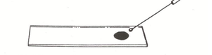
Place one drop of nigrosin on a clean slide.
Aseptically add a loopful of bacterial culture and mix gently.
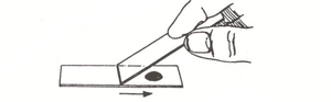
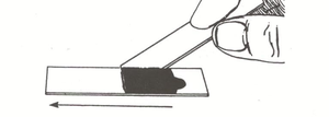
Use a second slide as a spreader: place its short edge at the drop and draw it into the drop.
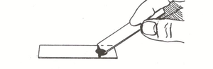
Allow the drop to spread across the edge of the spreader slide.
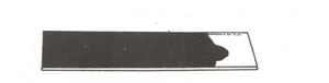
Draw the spreader slide toward the opposite end, spreading the stain.
Dispose of the spreader slide in a sharps container.
Air dry the slide; do not heat fix.
Observe under 1000X magnification.
Interpretation of Negative Stains
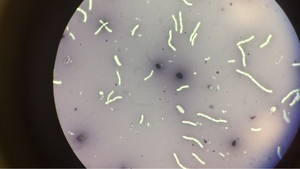
Negative stains allow clear visualization of cell shape and arrangement. Cells appear as clear, colorless objects against a dark background.
Morphology: Shapes such as cocci, bacilli, spirilla can be distinguished.
Arrangement: Cells may be found singly, in pairs, chains, or clusters.
Capsule Staining
Principles and Applications
Capsule staining is a differential technique used to visualize the capsule, a gelatinous layer surrounding some bacteria. Capsules are important for virulence and protection against host defenses. The procedure avoids heat fixation to prevent capsule distortion.
Purpose: To detect and visualize bacterial capsules.
Key Dyes: Crystal violet (primary stain), copper sulfate (decolorizer/secondary stain).
Medium: Bacteria are grown in protein-rich media (e.g., litmus milk) to enhance capsule formation.
Applications: Used for bacteria such as Enterobacter aerogenes and Staphylococcus epidermidis.
Capsule Stain Procedure
Aseptically add a loopful of culture grown in litmus milk to a slide and spread thinly.
Allow smear to air dry; do not heat fix.
Cover smear with 1% crystal violet for 2 minutes.
Rinse gently with 20% copper sulfate.
Air dry again; do not blot dry.
Observe under 1000X magnification.
Interpretation of Capsule Stains
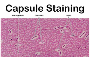
Capsule stains result in cells and background stained purple, while capsules appear as clear halos around cells. The copper sulfate removes excess crystal violet from the capsule, enhancing contrast.
Capsule: Clear, unstained area surrounding the cell.
Cell: Stained purple by crystal violet.
Background: Also stained purple.
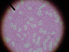
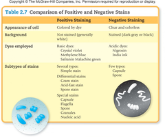
Comparison of Positive and Negative Staining
Key Differences
Positive and negative staining differ in their approach, dyes used, and the appearance of cells and background.
Feature | Positive Staining | Negative Staining |
|---|---|---|
Appearance of cell | Colored by dye | Clear and colorless |
Background | Unstained (generally white) | Stained (dark gray or black) |
Dyes employed | Basic dyes (crystal violet, methylene blue, safranin, malachite green) | Acidic dyes (nigrosin, India ink) |
Subtypes of stains | Several types: Differential stains (Gram, acid-fast, spore), Special stains (capsule, flagella, nucleic acid) | Few types: Capsule, spore |
*Additional info: The table above is based on Table 2.7 from a microbiology textbook, summarizing the main differences between positive and negative staining techniques.*
Safety and Disposal
Laboratory Practices
Proper disposal of slides and sharps is essential for laboratory safety. Slides used in staining procedures should be disposed of in designated sharps containers or stored in appropriate boxes.
Sharps container: Used for disposal of spreader slides and stained slides.
Blue box: Used for storage of slides if not immediately disposed.
Summary
Key Points
Negative staining is ideal for observing fragile cells without distortion.
Capsule staining allows visualization of bacterial capsules, important for pathogenicity.
Both techniques avoid heat fixation to preserve cell structures.
Understanding staining methods is crucial for accurate identification and study of microorganisms.
