 Back
Microscopy and Cell Structure in Microbiology: Study Notes
Study Guide - Smart Notes
 Tailored notes based on your materials, expanded with key definitions, examples, and context.
Microscopes and Measurement in Microbiology
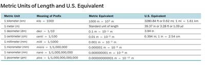
Units of Measurement for Microorganisms
Microorganisms are measured using the metric system, primarily in micrometers (μm) and nanometers (nm). Understanding these units is essential for interpreting microscopic observations and scientific literature.
Micrometer (μm): 1 μm = 10-6 meters (one-millionth of a meter).
Nanometer (nm): 1 nm = 10-9 meters (one-billionth of a meter).
Other units include millimeter (mm), centimeter (cm), and kilometer (km), but these are less commonly used for microorganisms.
Metric Unit | Meaning of Prefix | Metric Equivalent | U.S. Equivalent |
|---|---|---|---|
Kilometer (km) | kilo = 1,000 | 1,000 m = 103 m | 0.62 mi |
Meter (m) | Standard unit | 1 m | 39.37 in |
Centimeter (cm) | centi = 1/100 | 0.01 m = 10-2 m | 0.394 in |
Millimeter (mm) | milli = 1/1,000 | 0.001 m = 10-3 m | 0.039 in |
Micrometer (μm) | micro = 1/1,000,000 | 0.000001 m = 10-6 m | 0.000039 in |
Nanometer (nm) | nano = 1/1,000,000,000 | 0.000000001 m = 10-9 m | 0.000000039 in |
Picometer (pm) | pico = 1/1,000,000,000,000 | 0.000000000001 m = 10-12 m | 0.000000000039 in |
Principles and Types of Microscopy
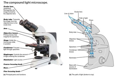
Compound Light Microscope: Structure and Light Path
The compound light microscope is the most common instrument in microbiology. It uses a series of lenses and visible light to magnify specimens.
Ocular lens (eyepiece): Magnifies the image formed by the objective lens, typically 10×.
Objective lenses: Closest to the specimen; common magnifications are 4×, 10×, 40×, and 100× (oil immersion).
Condenser: Focuses light onto the specimen.
Illuminator: Light source for the microscope.
Path of light: Illuminator → Condenser → Specimen → Objective lens → Body tube → Ocular lens → Eye.
Total Magnification and Resolution
Total magnification is calculated by multiplying the magnification of the objective lens by that of the ocular lens:
Common total magnifications: 100×, 400×, 1000×.
Resolution (resolving power): The ability to distinguish two points as separate entities. The shorter the wavelength of light, the greater the resolution.
Types of Light Microscopy
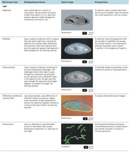
Different types of light microscopy are used to visualize microorganisms based on their properties and the desired information.
Microscope Type | Distinguishing Features | Principal Uses |
|---|---|---|
Brightfield | Uses visible light; specimen appears dark against a bright background. | To observe stained specimens and count microbes. |
Darkfield | Uses a special condenser; specimen appears light against a dark background. | To observe live, unstained specimens (e.g., spirochetes). |
Phase-contrast | Enhances differences in refractive index; internal structures are visible. | Detailed examination of living cells. |
Differential Interference Contrast (DIC) | Uses two beams of light and prisms; produces 3D, colored images. | To provide three-dimensional images. |
Fluorescence | Uses UV light; fluorescent dyes emit visible light. | To rapidly detect and identify microbes using fluorescent antibodies. |
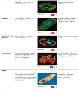
Confocal | Uses a single photon to illuminate one plane at a time. | To obtain two- and three-dimensional images of cells for biomedical applications. |
Two-photon | Uses two photons of red light to excite dyes. | To image living cells up to 1 mm deep. |
Super-Resolution Light Microscopy | Uses two laser beams to illuminate. | To observe locations of molecules in cells. |
Scanning Acoustic | Uses sound waves to produce images. | To study living cells attached to surfaces. |
Electron and Scanned-Probe Microscopy
Electron microscopes use electron beams instead of light, allowing much higher resolution and magnification. Scanned-probe microscopes use physical probes to map surfaces at the atomic level.
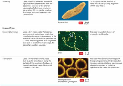
Transmission Electron Microscope (TEM): Electrons pass through ultrathin sections; used to study internal structures.
Scanning Electron Microscope (SEM): Electrons scan the surface; produces 3D images of surfaces.
Scanning Tunneling Microscope (STM): Uses a metal probe to scan the surface at atomic resolution.
Atomic Force Microscope (AFM): Uses a metal-and-diamond probe to produce 3D images of surfaces and molecules.
Staining Techniques in Microbiology
Simple, Differential, and Special Stains
Staining enhances contrast in microscopic images and allows for the identification and classification of microorganisms.
Simple stains: Use a single dye to highlight the entire microorganism.
Differential stains: Distinguish between different types of bacteria (e.g., Gram stain, acid-fast stain).
Special stains: Used to color specific structures (e.g., capsules, endospores, flagella).
Stain | Principal Uses |
|---|---|
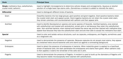
Simple | Highlight microorganisms to determine shapes and arrangements. |
Gram | Classifies bacteria as gram-positive or gram-negative. |
Acid-fast | Identifies Mycobacterium and Nocardia species. |
Negative | Demonstrates capsules as halos around cells. |
Endospore | Demonstrates presence of endospores in bacteria. |
Flagella | Demonstrates presence and arrangement of flagella. |
Gram Stain Procedure and Interpretation
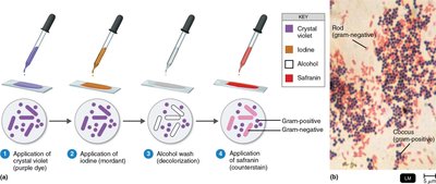
The Gram stain is a differential staining technique that classifies bacteria into two groups based on cell wall structure.
Application of crystal violet (primary stain) – all cells stain purple.
Application of iodine (mordant) – forms a complex with crystal violet.
Alcohol wash (decolorization) – removes stain from gram-negative cells.
Application of safranin (counterstain) – stains gram-negative cells pink.
Gram-positive bacteria: Retain crystal violet and appear purple.
Gram-negative bacteria: Lose crystal violet after alcohol wash and appear pink after safranin.
Prokaryotic Cell Structure and Function
Overview of Prokaryotic Cell Structure
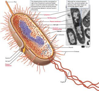
Prokaryotic cells, such as bacteria, are structurally simpler than eukaryotic cells. They lack a nucleus and membrane-bound organelles but contain essential components for life.
Cell wall: Provides shape and protection; contains peptidoglycan.
Plasma membrane: Selective barrier for transport.
Cytoplasm: Contains ribosomes, inclusions, and the nucleoid (DNA region).
External structures: Capsule, flagella, fimbriae, and pili.
Bacterial Shapes and Arrangements
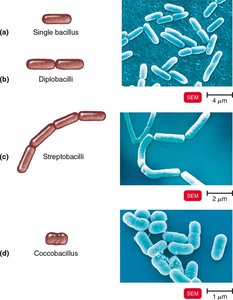
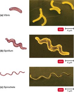
Bacteria exhibit a variety of shapes and arrangements, which are important for identification.
Coccus (spherical): Can form pairs (diplococci), chains (streptococci), clusters (staphylococci), tetrads, or sarcinae.
Bacillus (rod-shaped): Can be single, paired (diplobacilli), chained (streptobacilli), or coccobacilli (oval).
Spiral: Includes vibrios (curved rods), spirilla (rigid, helical), and spirochetes (flexible, helical).
Flagella, Axial Filaments, Fimbriae, and Pili
These structures are involved in motility, attachment, and genetic exchange.
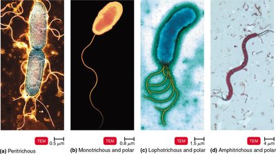
Flagella: Long, whip-like appendages for movement; arrangements include peritrichous, monotrichous, lophotrichous, and amphitrichous.
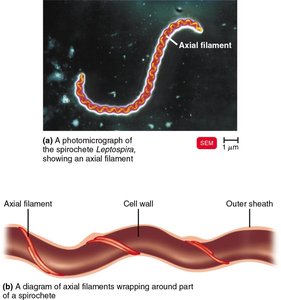
Axial filaments: Found in spirochetes; enable corkscrew movement.
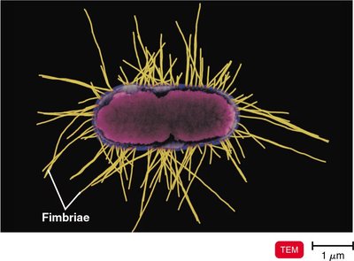
Fimbriae: Short, hair-like structures for attachment to surfaces and biofilm formation.
Pili: Longer than fimbriae; involved in motility (twitching, gliding) and DNA transfer (conjugation).
Gram-Positive vs. Gram-Negative Cell Walls
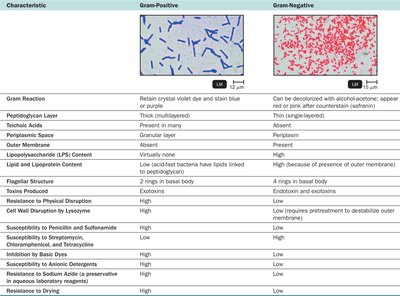
Bacterial cell walls differ in structure and chemical composition, influencing their staining properties and susceptibility to antibiotics.
Characteristic | Gram-Positive | Gram-Negative |
|---|---|---|
Peptidoglycan Layer | Thick (multilayered) | Thin (single-layered) |
Teichoic Acids | Present | Absent |
Outer Membrane | Absent | Present (contains LPS) |
Susceptibility to Penicillin | High | Low |
Resistance to Physical Disruption | High | Low |
Prokaryotic Ribosomes
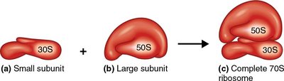
Ribosomes are the sites of protein synthesis. Prokaryotic ribosomes are 70S, composed of a 50S large subunit and a 30S small subunit.
Prokaryotic vs. Eukaryotic Cells: Summary Table
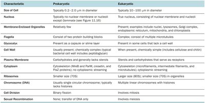
Characteristic | Prokaryotic | Eukaryotic |
|---|---|---|
Size | 0.2–2.0 μm | 10–100 μm |
Nucleus | Absent | Present |
Organelles | Absent | Present |
Cell Wall | Peptidoglycan (bacteria) | Cellulose, chitin, or none |
Ribosomes | 70S | 80S (70S in mitochondria/chloroplasts) |
Cell Division | Binary fission | Mitosis/meiosis |
Additional info: These notes cover essential microscopy and cell structure concepts for microbiology students, including measurement units, microscope types, staining techniques, and the structural differences between prokaryotic and eukaryotic cells. Understanding these foundations is critical for further study in microbial physiology, genetics, and pathogenesis.
