 Back
BackMicroscopy and Differential Staining Techniques in Microbiology
Study Guide - Smart Notes
 Tailored notes based on your materials, expanded with key definitions, examples, and context.
Microscopes
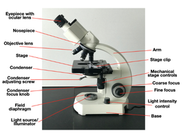
Parts of the Microscope and Their Functions
The compound light microscope is an essential tool in microbiology, allowing for the visualization of microorganisms too small to be seen with the naked eye. Understanding the parts and their functions is crucial for proper use and maintenance.
Eyepiece with ocular lens: The lens you look through, typically 10x magnification.
Nosepiece: Holds and rotates the objective lenses.
Objective lens: Provides primary magnification (commonly 4x, 10x, 40x, 100x).
Stage: Platform where the slide is placed.
Condenser: Focuses light onto the specimen.
Condenser adjusting screw: Moves the condenser up or down.
Condenser focus knob: Fine-tunes the focus of the condenser.
Field diaphragm: Controls the diameter of the light beam.
Light source/illuminator: Provides illumination for viewing specimens.
Arm: Supports the body tube and connects it to the base.
Stage clip: Holds the slide in place.
Mechanical stage controls: Move the slide on the stage.
Coarse focus: Brings the specimen into general focus.
Fine focus: Sharpens the focus for detailed viewing.
Light intensity control: Adjusts the brightness of the light source.
Base: Supports the microscope.
Proper Handling and Storage of the Microscope
Always carry the microscope with both hands: one on the arm and one under the base.
Clean lenses with lens paper only.
After use, lower the stage, set the lowest objective lens in place, and cover the microscope.
Total Magnification Calculation
Total magnification is determined by multiplying the ocular lens magnification by the objective lens magnification:
Formula:
Examples:
Ocular 10x × Objective 40x = 400x
Ocular 10x × Objective 100x = 1000x
Oil Immersion Technique
Oil immersion is used to increase the resolving power of the microscope when using the 100x objective lens.
Place a drop of immersion oil on the slide before rotating the 100x lens into position.
The oil has a refractive index similar to glass, reducing light refraction and allowing more light to enter the lens.
This results in a brighter, clearer image, essential for viewing bacteria and other small structures.
Microscopes
Types of Media in Microbiology
Different types of media are used to culture and study microorganisms, each with specific advantages:
TSA Plate (Tryptic Soy Agar): Large surface area, ideal for counting colonies and visualizing growth.
TSA Slant: Greater surface area in a tube, reduced risk of contamination and desiccation, easy to store and transport.
TSB Tube (Tryptic Soy Broth): Allows growth of large numbers of bacteria in a small volume.
Microscopes
Gram Staining
Gram staining is a differential staining technique used to classify bacteria based on cell wall structure. It distinguishes between Gram-positive and Gram-negative bacteria.
Crystal Violet (Primary Stain): Stains all cells purple.
Iodine (Mordant): Forms a complex with crystal violet, fixing it inside the cell wall.
Alcohol or Acetone (Decolorizer): Removes the crystal violet-iodine complex from Gram-negative cells only.
Safranin (Counterstain): Stains decolorized Gram-negative cells pink/red; Gram-positive cells remain purple.
Structural Differences Between Gram-Positive and Gram-Negative Bacteria
The main difference lies in the cell wall composition:
Gram-positive: Thick peptidoglycan layer, no outer membrane.
Gram-negative: Thin peptidoglycan layer, outer membrane with lipopolysaccharides.
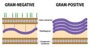
Gram Stain Results
Gram-positive bacteria: Appear purple (e.g., Staphylococcus epidermidis).
Gram-negative bacteria: Appear pink/red (e.g., Escherichia coli).
Microscopes
Acid-Fast Stain
The acid-fast stain is used to identify bacteria with waxy cell walls, such as Mycobacterium species.
Carbol Fuchsin (Primary Stain): Stains all cells red; acid-fast cells retain this color due to their waxy cell wall.
Acid-Alcohol (Decolorizer): Removes stain from non-acid-fast cells; acid-fast cells remain red.
Methylene Blue (Counterstain): Stains non-acid-fast cells blue for contrast.
Positive result: Red (acid-fast bacteria).
Negative result: Blue (non-acid-fast bacteria).
Acid-fast staining is crucial for diagnosing diseases like tuberculosis. The key cell wall component is mycolic acid, a waxy, long-chain fatty acid that makes the cell wall impermeable to most stains.
Microscopes
Endospore Stain
The endospore stain differentiates endospores from vegetative cells, important for identifying spore-forming bacteria.
Malachite Green (Primary Stain): Stains endospores green; heat is used to facilitate dye penetration.
Water (Decolorizer): Removes stain from vegetative cells but not from endospores.
Safranin (Counterstain): Stains vegetative cells red or pink, providing contrast to green endospores.
Positive result: Green endospores.
Negative result: Red or pink vegetative cells.
Bacterial genera that form endospores include Bacillus (e.g., Bacillus anthracis) and Clostridium (e.g., Clostridium botulinum, Clostridium tetani). Only some Gram-positive bacteria can form endospores.
Bacteria form endospores in response to adverse conditions such as:
Nutrient depletion (especially carbon or nitrogen)
Extreme temperatures
Desiccation
Radiation
Exposure to toxic chemicals
These conditions trigger bacteria to enter a dormant, highly resistant state by forming endospores.
