Back
Microscopy and Prokaryotic Cell Morphology in Microbiology
Study Guide - Smart Notes
 Tailored notes based on your materials, expanded with key definitions, examples, and context.
Microscopy in Microbiology
Types of Microscopes
Microscopes are essential tools in microbiology, allowing for the visualization of microorganisms and cellular structures that are invisible to the naked eye. There are three main classes of microscopes, each with unique mechanisms and applications:
Light Microscopes: Use visible or ultraviolet light to illuminate specimens. Types include:
Bright-field
Dark-field
Fluorescence
Confocal
Electron Microscopes: Use beams of electrons for much higher resolution and magnification. Types include:
Transmission Electron Microscope (TEM)
Scanning Electron Microscope (SEM)
Scanning Probe Microscopes: Use physical probes to scan the surface of specimens, such as:
Atomic Force Microscope (AFM)
Scanning Tunneling Microscope

Principles of Microscopy
Microscopy relies on several physical principles to generate images of small objects:
Wavelength of Radiation: Shorter wavelengths provide greater resolving power, allowing for the visualization of smaller structures.
Refraction and Magnification: Refraction is the bending of light as it passes through different media, while magnification is the perceived enlargement of the image. The total magnification is calculated as:
Resolution: The ability to distinguish two adjacent objects as separate entities.
Contrast: The difference in intensity between an object and its background, often enhanced by staining.
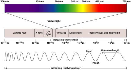
Bright-Field Microscopy
Bright-field microscopes are the most common type used in microbiology. They use a series of lenses to magnify the specimen, which appears dark against a bright background. Oil immersion lenses are used to increase resolution at high magnifications.
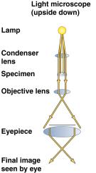
Other Forms of Light Microscopy
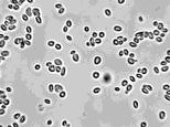
Dark-Field Microscopy: Increases contrast by making the specimen appear light against a dark background, useful for observing live, unstained specimens.
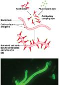
Fluorescence Microscopy: Uses high-intensity UV light to excite fluorescent molecules in the specimen, which then emit visible light. This technique is valuable for identifying specific microbes using fluorescent dyes or antibodies.
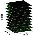
Confocal Microscopy: Uses lasers to scan specimens stained with fluorescent dyes, producing sharp, three-dimensional images by stacking optical sections.
Electron Microscopy
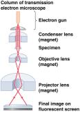
Electron microscopes use electron beams instead of light, providing much higher resolution and magnification. They are essential for visualizing viruses, internal cell structures, and large molecules.
Transmission Electron Microscope (TEM): Electrons pass through thin sections of the specimen, revealing internal structures.
Scanning Electron Microscope (SEM): Electrons scan the surface, producing detailed three-dimensional images of specimen surfaces.
Scanning Probe Microscopy
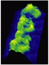
Scanning probe microscopes, such as AFM, use a physical probe to scan the surface of a specimen, allowing visualization of single molecules and atomic clusters with extremely high magnification.
Staining Techniques in Microbiology
Purpose and Principles of Staining
Staining increases contrast and resolution by coloring specimens with dyes. Most bacterial cells have a net negative charge, so basic dyes (positively charged) are commonly used. Stains are usually salts with a colored ion (chromophore).
Acidic Dyes: Stain alkaline structures (e.g., Indian ink).
Basic Dyes: Stain acidic structures (e.g., crystal violet).
Smear Preparation: Microorganisms are spread in a thin film, air-dried, and fixed to the slide by heat or chemicals.
Types of Stains
Simple Stains: Use a single dye to color all cells, revealing cell shape and arrangement.
Differential Stains: Use multiple dyes to distinguish between different types of cells or structures. Examples include:
Gram Stain
Acid-Fast Stain
Endospore Stain
Special Stains: Highlight specific structures, such as capsules or flagella.
Gram Stain
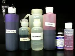
The Gram stain is a four-step differential stain that classifies bacteria based on cell wall composition:
Crystal violet (primary stain)
Iodine (mordant)
Alcohol (decolorizer)
Safranin (counterstain)
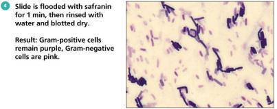
Results: Gram-positive bacteria stain purple; Gram-negative bacteria stain pink. This distinction is crucial for bacterial identification and antibiotic selection.
Prokaryotic Cell Morphology and Arrangements
Shapes of Prokaryotic Cells
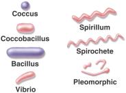
Prokaryotic cells exhibit a variety of shapes, which are important for classification and identification:
Coccus (spherical)
Bacillus (rod-shaped)
Vibrio (curved rod)
Spirillum (rigid spiral)
Pleomorphic (variable shape)
Arrangements of Prokaryotic Cells
Cell arrangements result from the plane of cell division and whether daughter cells separate:
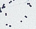
Cocci Arrangements:
Diplococci (pairs)
Streptococci (chains)
Staphylococci (clusters)
Tetrads (groups of four)
Sarcina (cubical packets)
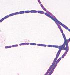
Bacilli Arrangements:
Single bacilli
Diplobacilli (pairs)
Streptobacilli (chains)
V-shaped and palisade (side-by-side)
Reproduction of Prokaryotic Cells
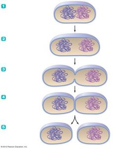
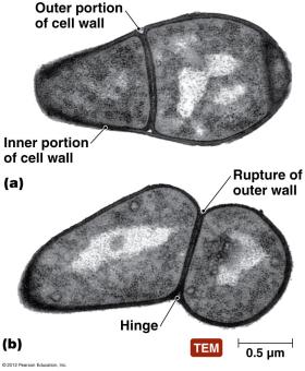
All prokaryotes reproduce asexually, most commonly by binary fission. Some Gram-positive bacteria use a variation called snapping division.
Binary Fission: The cell replicates its DNA, elongates, forms a cross wall, and divides into two daughter cells.
Snapping Division: The inner cell wall grows and creates tension, causing the outer wall to snap and the cells to remain attached.
Endospores
Structure and Function of Endospores
Endospores are specialized, highly resistant structures produced by certain Gram-positive bacteria (e.g., Bacillus and Clostridium species). They are not reproductive structures but serve as a survival mechanism under harsh conditions.
Each vegetative cell forms one endospore, which can germinate into a new cell.
Endospores are extremely resistant to desiccation, heat, radiation, and chemicals, making them difficult to eradicate.
Endospore formation is a concern in food safety and healthcare due to their persistence and resistance.
Endospore Formation (Sporulation)
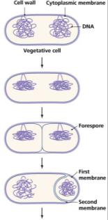
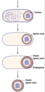
The process of endospore formation involves several steps:
DNA replication
Formation of a forespore
Development of protective layers (cortex and spore coats)
Maturation and release of the endospore
Examples of Endospores
Medial Endospores: Located centrally within the cell (e.g., Bacillus species).
Terminal Endospores: Located at the end of the cell (e.g., Clostridium species).