 Back
Microscopy and Prokaryotic Cell Structure: Foundations for Microbiology
Study Guide - Smart Notes
 Tailored notes based on your materials, expanded with key definitions, examples, and context.
Microscopy
Principles of Light Microscopy
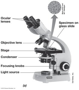
Microscopy is a fundamental technique in microbiology, allowing visualization of microorganisms and their structures. The compound light microscope is the most common tool, utilizing two sets of lenses (ocular and objective) and visible light to magnify specimens.
Ocular lenses: The eyepieces through which the specimen is viewed.
Objective lenses: Provide primary magnification; several objectives of varying power are typically available.
Stage: Platform where the specimen slide is placed.
Condenser: Focuses light onto the specimen.
Focusing knobs: Adjust the focus for clear visualization.
Light source: Illuminates the specimen from below.
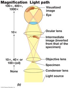
Magnification is the product of the ocular and objective lens powers (e.g., 10× ocular × 100× objective = 1000× total magnification). Resolution is the minimum distance at which two points can be distinguished, typically about 0.2 μm for light microscopes. Resolution depends on the wavelength of light and the numerical aperture of the lens.
Types of Light Microscopy
Different light microscopy techniques enhance contrast and reveal various cellular features:
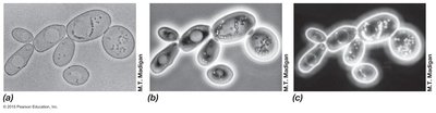
Bright-field microscopy: Relies on light absorption or scattering by the specimen to create contrast.
Phase-contrast microscopy: Exploits differences in refractive index between cell components and surroundings, enhancing visualization of transparent specimens.
Dark-field microscopy: Illuminates the specimen from the side, so only scattered light enters the objective, making specimens appear bright against a dark background.
Fluorescence and Advanced Light Microscopy
Fluorescence microscopy uses fluorescent dyes or proteins to label specific cellular components, allowing visualization of structures with high specificity. Multiple stains can be used to distinguish different targets simultaneously.
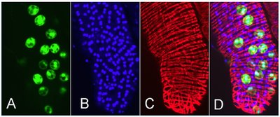
Advanced 3D light microscopy techniques include:
Differential Interference Contrast (DIC): Uses polarized light to enhance contrast and produce pseudo-3D images.
Confocal Laser Scanning Microscopy (CLSM): Uses lasers and computer assembly of optical sections to create detailed 3D reconstructions, ideal for thick specimens.
Electron Microscopy
Electron microscopes use electron beams instead of light, achieving much higher resolution (up to 1000× better than light microscopy), allowing visualization of viruses and molecular structures.
Transmission Electron Microscopy (TEM): Electrons pass through thin sections of specimens, revealing internal structures.
Scanning Electron Microscopy (SEM): Electrons scan the specimen surface, producing detailed 3D images of external features.
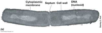
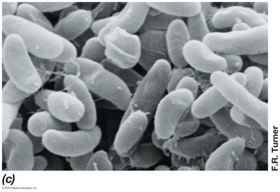
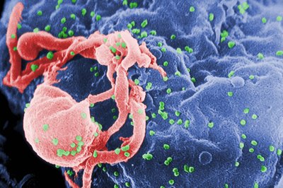
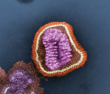
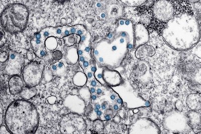
Electron micrographs are often false-colored to highlight specific features or organisms.
Prokaryotic Cell Structure and Morphology
Major Cell Morphologies
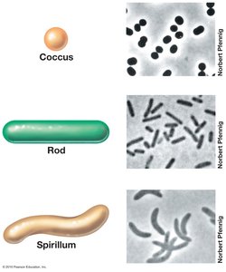
Prokaryotes exhibit a variety of cell shapes, which are important for identification and ecological function:
Coccus: Spherical shape
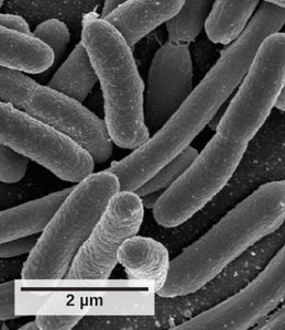
Rod (Bacillus): Cylindrical shape
Spirillum: Spiral-shaped
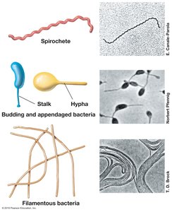
Unusual morphologies include square-shaped Haloquadratum and filamentous or appendaged bacteria.
Dynamic and Large Prokaryotic Cells
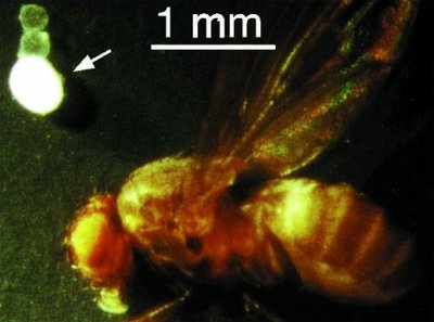
Some prokaryotes can change their morphology in response to environmental conditions (e.g., Helicobacter pylori, Myxococcus). Exceptionally large prokaryotes include Epulopiscium fishelsoni and Thiomargarita namibiensis, which can be hundreds of micrometers in size.
Advantages of Small Cell Size
Most prokaryotes are small, which provides several advantages:
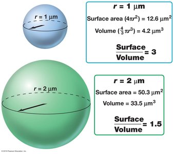
High surface area-to-volume (S/V) ratio enables rapid nutrient uptake and waste removal.
Small cells can be produced in greater numbers, supporting large populations and rapid adaptation.
The S/V ratio is calculated as:
Microbial Membranes and Cell Walls
Cytoplasmic Membrane Structure
The cytoplasmic membrane is a selectively permeable barrier that encloses the cell contents. It is composed of a lipid bilayer with embedded proteins. Bacterial and eukaryotic membranes contain ester-linked phospholipids, while archaeal membranes have ether-linked lipids and may form monolayers, especially in hyperthermophiles.
Selective permeability: Allows certain substances to pass while restricting others.
Protein anchor: Membrane proteins facilitate transport and energy generation.
Membrane Permeability and Transport
Different substances cross the membrane at different rates. Small, uncharged molecules like water diffuse easily, while ions and large molecules require transport proteins.
Substance | Relative Permeability | Diffusion into Cell |
|---|---|---|
Water | 100 | Excellent |
Glycerol | 0.1 | Good |
Tryptophan | 0.001 | Fair/Poor |
Glucose | 0.001 | Fair/Poor |
Chloride ion (Cl−) | 0.000001 | Very Poor |
Potassium ion (K+) | 0.0000001 | Extremely Poor |
Sodium ion (Na+) | 0.00000001 | Extremely Poor |
Transport proteins enable cells to accumulate essential nutrients from dilute environments, supporting survival in nutrient-poor conditions.
Gram-Positive vs. Gram-Negative Cell Walls
Prokaryotic cell walls provide structural support and protection. The Gram stain differentiates bacteria based on cell wall structure:
Gram-positive (G+): Thick peptidoglycan layer retains crystal violet stain.
Gram-negative (G−): Thin peptidoglycan layer and an outer membrane containing lipopolysaccharides (LPS), which can be toxic in pathogens.
Summary Table: Key Differences in Prokaryotic Cell Envelopes
Feature | Gram-Positive | Gram-Negative | Archaea |
|---|---|---|---|
Peptidoglycan Layer | Thick | Thin | Absent (pseudopeptidoglycan or S-layer) |
Outer Membrane | Absent | Present (LPS) | Absent |
Lipid Linkage | Ester | Ester | Ether |
Typical Stain Reaction | Purple | Pink | Variable |
Example: Pelagibacter ubique (SAR11) is a small, abundant marine bacterium with a minimal genome, illustrating the efficiency and adaptability of prokaryotic cell structure.
Additional info: Advanced microscopy techniques and cell envelope diversity are foundational for understanding microbial physiology, ecology, and taxonomy.
