 Back
BackMicroscopy and Staining in Microbiology
Study Guide - Smart Notes
 Tailored notes based on your materials, expanded with key definitions, examples, and context.
Microscopy and Staining
Introduction to Microscopy in Microbiology
Microscopy is a fundamental technique in microbiology, allowing scientists to observe organisms and structures too small to be seen with the naked eye. The choice of microscope and staining method depends on the size, structure, and properties of the specimen being studied.
Microscopy
Scale of Biological Structures
Understanding the relative sizes of biological structures is essential for selecting the appropriate microscopy technique. Microorganisms range from the size of eukaryotic cells (about 10–100 μm) to viruses (about 0.01–0.1 μm), which are much smaller than most cells and organelles.
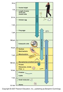
Eukaryotic cells: 10–100 μm
Most bacteria: 1–10 μm
Viruses: 0.01–0.1 μm
Proteins and lipids: 1–10 nm
Additional info: The resolving power of a microscope must match the size of the specimen for effective visualization.
Parts and Function of the Light Microscope
The compound light microscope is the most commonly used instrument in microbiology. It consists of several key components that work together to magnify and resolve the image of a specimen.
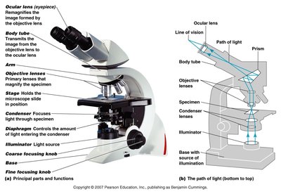
Ocular lens (eyepiece): Remagnifies the image formed by the objective lens.
Objective lenses: Primary lenses that magnify the specimen (typically 4x, 10x, 40x, 100x).
Stage: Holds the microscope slide in position.
Condenser: Focuses light through the specimen.
Illuminator: Light source for the microscope.
Coarse and fine focusing knobs: Adjust the focus of the image.
Oil Immersion Technique
Oil immersion is used with high-power objective lenses (usually 100x) to increase resolution by reducing light refraction. Immersion oil has a similar refractive index to glass, allowing more light to enter the objective lens.
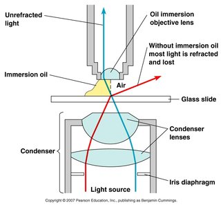
Without oil: Light is refracted and lost, reducing image clarity.
With oil: More light passes directly into the objective lens, improving resolution.
Types of Light Microscopy
Different types of light microscopy are used to visualize specimens based on their properties and the desired information.
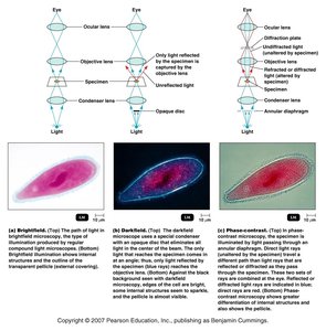
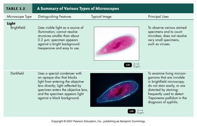
Brightfield microscopy: Illuminates the specimen directly; best for stained, fixed specimens.
Darkfield microscopy: Uses an opaque disc to block direct light, making specimens appear bright against a dark background; useful for live, unstained organisms.
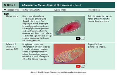
Phase-contrast microscopy: Enhances contrast in transparent specimens by amplifying differences in refractive index; ideal for observing living cells and internal structures.
Summary Table: Types of Microscopes
The following table summarizes the main types of microscopes, their distinguishing features, typical images, and principal uses.
Microscope Type | Distinguishing Features | Typical Image | Principal Uses |
|---|---|---|---|
Brightfield | Uses visible light as a source of illumination; contrast reveals structures smaller than about 0.2 μm; specimen appears against a bright background. |
| To observe various stained specimens and count microbes; does not resolve very small specimens, such as viruses. |
Darkfield | Uses a special condenser with an opaque disc; only light reflected by the specimen enters the objective lens; specimen appears bright against a dark background. |
| To examine living microorganisms that are invisible in brightfield microscopy or cannot be stained easily. |
Phase-contrast | Uses a special condenser containing an annular (ring-shaped) diaphragm; enhances contrast of transparent specimens without staining. |
| To facilitate detailed examination of internal structures of living specimens. |
Differential interference contrast (DIC) | Like phase-contrast, uses differences in refractive indexes to produce images; two beams of light separated by prisms create a three-dimensional effect. |
| To provide three-dimensional images. |
Fluorescence | Uses ultraviolet or near-ultraviolet light to cause fluorescent microbes to emit light. |
| For fluorescent-antibody techniques to rapidly detect and identify microbes in tissues or clinical specimens. |
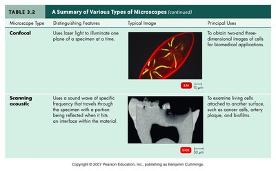
Confocal | Uses laser light to illuminate one plane of a specimen at a time. |
| To obtain three-dimensional images of cells for biomedical applications. |
Scanning acoustic | Uses sound waves of specific frequency that travel through the specimen; reflected when hitting an interface within the material. |
| To examine living cells attached to another surface, such as cancer cells, artery plaque, and biofilms. |
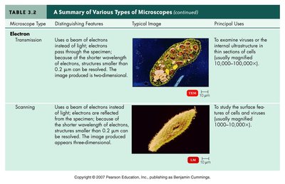
Transmission electron (TEM) | Uses a beam of electrons instead of light; electrons pass through the specimen, revealing structures smaller than 0.2 μm; image is two-dimensional. |
| To examine viruses or the internal structure of thin sections of cells. |
Scanning electron (SEM) | Uses a beam of electrons reflected off the surface of the specimen; produces a three-dimensional image. |
| To study the surface features of cells and viruses. |
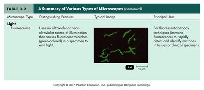
Fluorescence Microscopy and Immunofluorescence
Fluorescence microscopy uses ultraviolet light to excite fluorescent molecules in the specimen, causing them to emit visible light. Immunofluorescence is a technique where antibodies are labeled with fluorescent dyes to detect specific antigens in microorganisms.
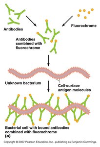
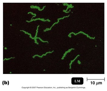
Application: Rapid identification of pathogens in clinical samples.
Electron Microscopy
Electron microscopes use beams of electrons instead of light, allowing for much higher resolution and magnification. There are two main types: transmission electron microscopy (TEM) and scanning electron microscopy (SEM).
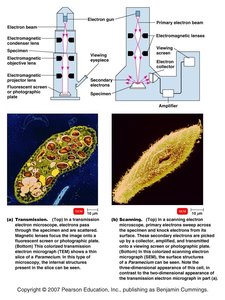
TEM: Electrons pass through thin sections of the specimen, revealing internal structures in high detail.
SEM: Electrons scan the surface, producing detailed three-dimensional images of specimen surfaces.
Staining Microbes
Purpose and Principles of Staining
Staining enhances the contrast of microscopic specimens, making cellular structures more visible. Different stains and staining techniques are used to distinguish between types of microorganisms and cellular components.
Gram Stain: Principle and Procedure
The Gram stain is a differential staining technique that classifies bacteria into two groups: Gram-positive and Gram-negative, based on differences in cell wall structure.
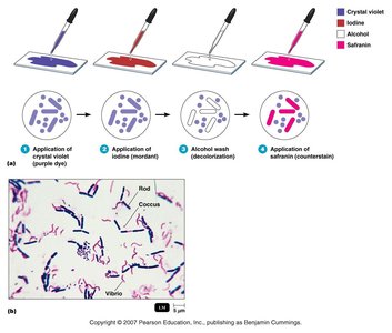
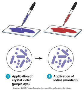
Step 1: Application of crystal violet (primary stain)
Step 2: Application of iodine (mordant)
Step 3: Alcohol wash (decolorization)
Step 4: Application of safranin (counterstain)
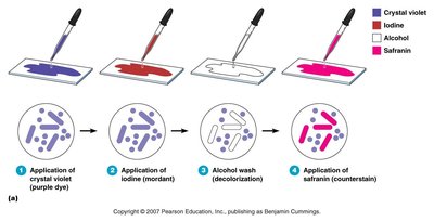
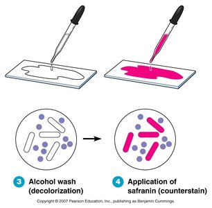
Result: Gram-positive bacteria retain the crystal violet stain (purple), while Gram-negative bacteria take up the safranin counterstain (pink/red).
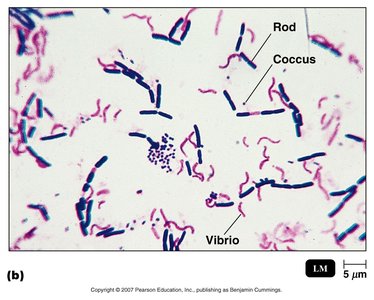
Gram-positive: Thick peptidoglycan layer, stains purple.
Gram-negative: Thin peptidoglycan layer, stains pink/red.
Other Staining Techniques
Special stains are used to visualize specific structures or types of microorganisms.
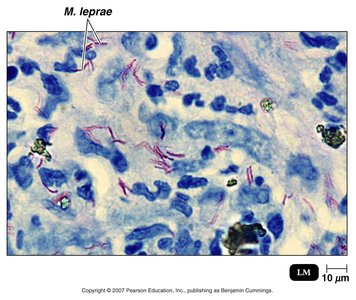
Acid-fast stain: Identifies bacteria with waxy cell walls, such as Mycobacterium species.
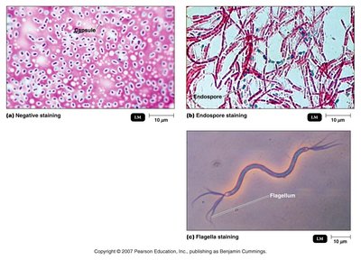
Negative stain: Visualizes capsules by staining the background, leaving the capsule clear.
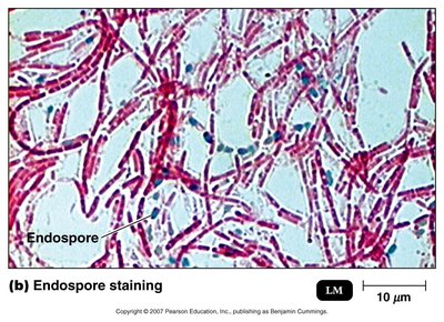
Endospore stain: Highlights bacterial endospores, which are resistant to harsh conditions.
Flagella stain: Makes bacterial flagella visible under the microscope.

Additional info: Staining is essential for microbial identification, classification, and understanding cellular structure and function.
