 Back
BackMicroscopy and Staining: Principles and Applications in Microbiology
Study Guide - Smart Notes
 Tailored notes based on your materials, expanded with key definitions, examples, and context.
Microscopy in Microbiology
Principles of Microscopy
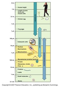
Microscopy is fundamental to microbiology, enabling visualization of organisms and structures too small to be seen with the naked eye. The scale of biological structures ranges from atoms and small molecules to cells and multicellular organisms, requiring different types of microscopes for observation.
Resolution: The ability to distinguish two points as separate entities. Higher resolution allows for more detailed observation.
Magnification: The process of enlarging the appearance of an object.
Types of Microscopes: Light microscopes (brightfield, darkfield, phase-contrast, fluorescence, confocal) and electron microscopes (transmission, scanning).
Applications: Used to study cell structure, morphology, and microbial classification.
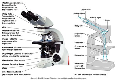
Structure and Function of the Light Microscope
The compound light microscope is the most commonly used instrument in microbiology. It consists of several key components that work together to magnify and resolve specimens.
Ocular lens (eyepiece): Remagnifies the image formed by the objective lens.
Objective lenses: Primary lenses that magnify the specimen.
Stage: Holds the microscope slide.
Condenser: Focuses light through the specimen.
Diaphragm: Controls the amount of light entering the condenser.
Illuminator: Light source.
Coarse and fine focusing knobs: Adjust the focus.
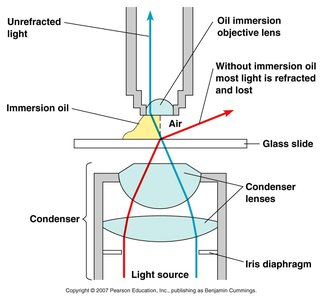
Oil Immersion Technique
Oil immersion is used to increase the resolution of the microscope when observing specimens at high magnification. Immersion oil reduces light refraction, allowing more light to enter the objective lens.
Without oil: Light is refracted and lost, reducing image clarity.
With oil: Light passes directly into the lens, improving resolution.
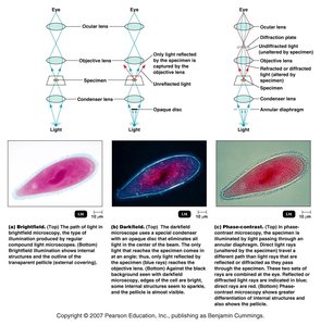
Types of Light Microscopy
Different light microscopy techniques are used to enhance contrast and reveal specific features of microorganisms.
Brightfield: Uses visible light; specimens appear dark against a bright background.
Darkfield: Uses an opaque disk to block direct light; specimens appear bright against a dark background.
Phase-contrast: Enhances contrast by amplifying differences in refractive index; useful for observing living cells.
Summary Table: Types of Microscopes
The following tables summarize the distinguishing features, typical images, and principal uses of various microscopes.
Microscope Type | Distinguishing Features | Typical Image | Principal Uses |
|---|---|---|---|
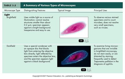
Brightfield | Uses visible light as a source of illumination; contrast reveals structures smaller than about 0.2 μm. |
| To observe various stained specimens and count microbes. |
Darkfield | Uses a special condenser with an opaque disc; light reflected by specimen enters objective lens. |
| To examine living microorganisms that are invisible in brightfield microscopy. |
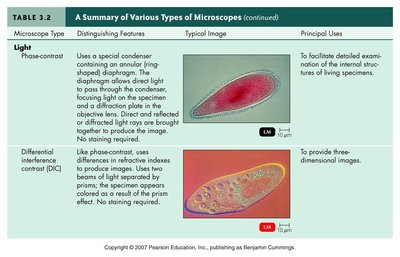
Phase-contrast | Uses a special condenser containing an annular diaphragm; enhances contrast. |
| To facilitate detailed examination of internal structures of living specimens. |
Differential Interference Contrast (DIC) | Uses differences in refractive indexes to produce images; two beams of light separated by prisms. |
| To provide three-dimensional images. |
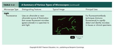
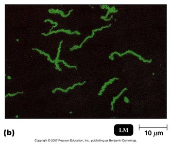
Fluorescence | Uses ultraviolet or near-ultraviolet source of illumination; causes fluorescent microbes to emit light. |
| For fluorescent-antibody techniques to rapidly detect and identify microbes. |
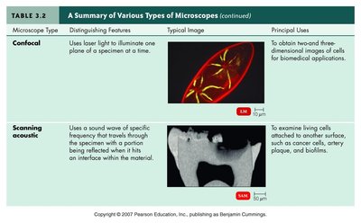
Confocal | Uses laser light to illuminate one plane of a specimen at a time. |
| To obtain three-dimensional images of cells for biomedical applications. |
Scanning Acoustic | Uses sound waves of specific frequency; examines living cells attached to surfaces. |
| To examine living cells attached to surfaces, such as cancer cells and biofilms. |
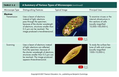
Transmission Electron (TEM) | Uses a beam of electrons instead of light; electrons pass through specimen. |
| To examine viruses or internal structures in thin sections of cells. |
Scanning Electron (SEM) | Uses a beam of electrons instead of light; electrons are reflected from specimen. |
| To study surface features of cells and viruses. |
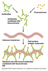
Fluorescence Microscopy and Immunofluorescence
Fluorescence microscopy uses fluorescent dyes or proteins to visualize specific structures or organisms. Immunofluorescence employs antibodies tagged with fluorochromes to detect antigens on microbial cells.
Antibodies: Proteins that bind specifically to antigens.
Fluorochrome: A fluorescent dye attached to antibodies.
Application: Used for rapid identification of pathogens in clinical specimens.
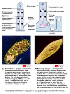
Electron Microscopy
Electron microscopes use beams of electrons instead of light, allowing for much higher resolution and magnification. Two main types are transmission electron microscopes (TEM) and scanning electron microscopes (SEM).
TEM: Electrons pass through thin sections of specimens, revealing internal structures.
SEM: Electrons are reflected from the surface, providing detailed three-dimensional images.
Staining Techniques in Microbiology
Principles of Staining
Staining is essential for visualizing and differentiating microorganisms under the microscope. It enhances contrast and allows for identification of cellular structures and classification of microbes.
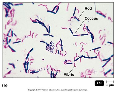
Simple Stain: Uses a single dye to color cells, making them easier to see.
Differential Stain: Uses multiple dyes to distinguish between different types of organisms or structures.
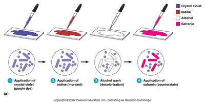
Gram Staining Procedure
Gram staining is a differential staining technique that classifies bacteria as Gram-positive or Gram-negative based on cell wall properties.
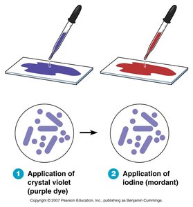
Application of crystal violet (primary dye)
Application of iodine (mordant)
Alcohol wash (decolorization)
Application of safranin (counterstain)
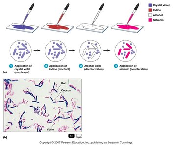
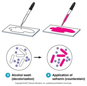
Gram-positive: Retain crystal violet and appear purple.
Gram-negative: Lose crystal violet, take up safranin, and appear pink/red.
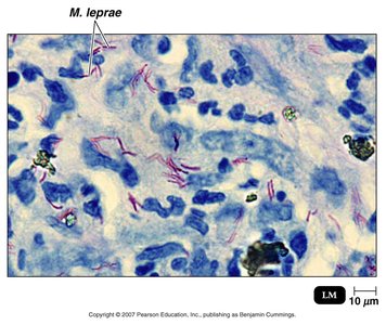
Acid-Fast Staining
Acid-fast staining is used to identify bacteria with waxy cell walls, such as Mycobacterium species.
Acid-fast bacteria: Retain the primary stain even after acid-alcohol wash.
Non-acid-fast bacteria: Lose the primary stain and take up the counterstain.
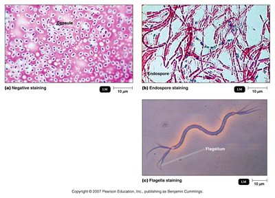
Special Staining Techniques
Special stains are used to highlight specific structures such as capsules, endospores, and flagella.
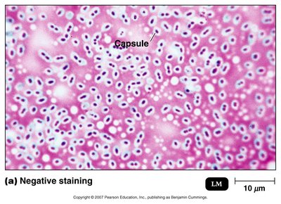
Negative staining: Stains the background, leaving capsules unstained.
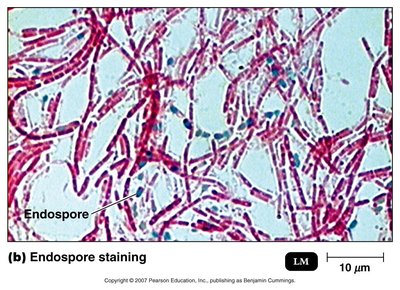
Endospore staining: Highlights endospores within bacterial cells.
Flagella staining: Makes flagella visible under the microscope.
Summary Table: Staining Techniques
Staining Technique | Purpose | Key Features |
|---|---|---|
Simple Stain | Visualize cell shape and arrangement | Uses one dye; all cells appear same color |
Gram Stain | Differentiate Gram-positive and Gram-negative bacteria | Uses crystal violet, iodine, alcohol, safranin |
Acid-Fast Stain | Identify acid-fast bacteria (e.g., Mycobacterium) | Uses carbol fuchsin, acid-alcohol, methylene blue |
Negative Stain | Visualize capsules | Stains background, not cells |
Endospore Stain | Visualize endospores | Uses malachite green and safranin |
Flagella Stain | Visualize flagella | Uses mordants to increase thickness |
Conclusion
Microscopy and staining are indispensable tools in microbiology, allowing for the visualization, differentiation, and classification of microorganisms. Mastery of these techniques is essential for understanding microbial structure, function, and diversity.
