 Back
Microscopy, Cell Domains, Staining, and Viruses: Microbiology Study Guide
Study Guide - Smart Notes
 Tailored notes based on your materials, expanded with key definitions, examples, and context.
Q1. Labeling the Parts of a Microscope and Understanding Magnification
Background
Topic: Microscopy Basics
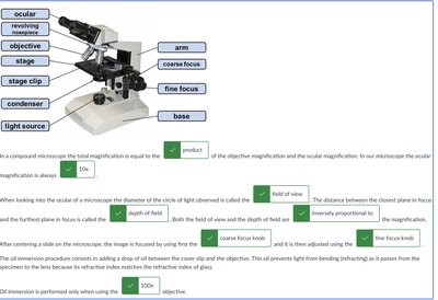
This question tests your knowledge of microscope parts, how total magnification is calculated, and the relationship between magnification, field of view, and depth of field.
Key Terms and Formulas:
Total Magnification:
Field of View: The diameter of the circle of light you see when looking into a microscope.
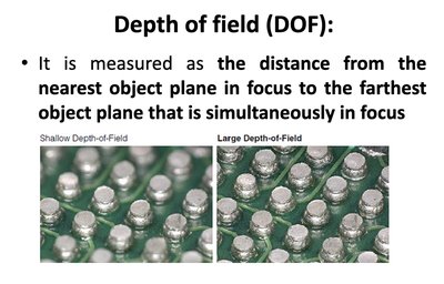
Depth of Field: The distance between the nearest and farthest objects in focus at one time.
Step-by-Step Guidance
Identify the labeled parts of the microscope (e.g., ocular, objective, stage, condenser, etc.).
Recall that total magnification is found by multiplying the ocular lens magnification by the objective lens magnification.
Understand that as you increase magnification, the field of view and depth of field both decrease.
Know which focus knob (coarse or fine) and which objective (lowest power) to use first when starting to view a specimen.
Be able to explain why oil immersion is used at high magnification (1000x) and how it improves image clarity.
Try solving on your own before revealing the answer!
Final Answer:
The total magnification is the product of the objective and ocular magnifications. As magnification increases, both the field of view and depth of field decrease. Start with the coarse focus knob and the lowest power objective. Oil immersion is used at 1000x to reduce light refraction and improve resolution.
Q2. Domains of Life: Bacteria, Archaea, and Eukarya
Background
Topic: Classification of Life
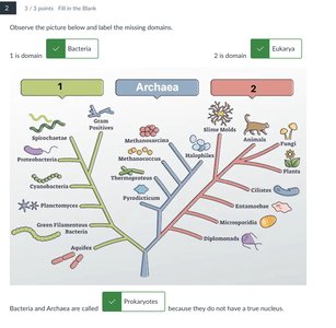
This question tests your understanding of the three domains of life and the differences between prokaryotes and eukaryotes.
Key Terms:
Bacteria: Prokaryotic domain, no true nucleus.
Archaea: Prokaryotic domain, no true nucleus, distinct from bacteria.
Eukarya: Domain with true nucleus (eukaryotes).
Prokaryotes: Organisms without a true nucleus (includes Bacteria and Archaea).
Step-by-Step Guidance
Identify the three domains of life: Bacteria, Archaea, and Eukarya.
Recognize that Bacteria and Archaea are both prokaryotes (no true nucleus), while Eukarya are eukaryotes (have a true nucleus).
Be able to label diagrams showing the three domains and classify organisms accordingly.
Understand the evolutionary relationships and differences between these domains.
Try solving on your own before revealing the answer!
Final Answer:
Bacteria and Archaea are prokaryotes (no true nucleus), while Eukarya are eukaryotes (have a true nucleus). The three domains are Bacteria, Archaea, and Eukarya.
Q3. Field of View and Magnification in Microscopy
Background
Topic: Microscopy - Field of View
This question tests your understanding of how the field of view changes with magnification and how to measure it.
Key Terms and Concepts:
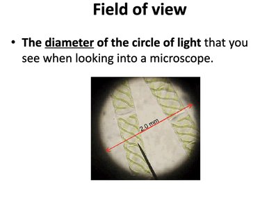
Field of View (FOV): The diameter of the visible area when looking through the microscope.
Magnification: The process of enlarging the appearance of an object.
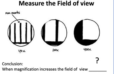
Relationship: As magnification increases, the field of view decreases.
Step-by-Step Guidance
Observe how the field of view is measured as the diameter of the circle of light seen through the microscope.
Compare the field of view at different magnifications (e.g., 40x, 100x, 400x) using diagrams or images.
Note the trend: as you increase magnification, the field of view becomes smaller.
Be able to explain why this happens in terms of optics and image enlargement.
Try solving on your own before revealing the answer!
Final Answer:
When magnification increases, the field of view decreases. This is because higher magnification zooms in on a smaller area of the specimen.
Q4. Depth of Field in Microscopy
Background
Topic: Microscopy - Depth of Field
This question tests your understanding of what depth of field means and how it changes with magnification.
Key Terms:
Depth of Field (DOF): The distance from the nearest object plane in focus to the farthest object plane that is simultaneously in focus.
Shallow vs. Large DOF: Shallow means only a thin layer is in focus; large means a thicker section is in focus.
Step-by-Step Guidance
Understand that depth of field is the range of depth that appears sharp in the microscope image.
Recognize that as magnification increases, the depth of field decreases (becomes shallower).
Be able to compare images with shallow and large depth of field and explain the differences.
Relate this concept to focusing techniques when using a microscope.
Try solving on your own before revealing the answer!
Final Answer:
Depth of field is the distance between the nearest and farthest objects in focus. As magnification increases, the depth of field decreases, making it harder to keep thick specimens in focus at high power.
Q5. Oil Immersion in Microscopy
Background
Topic: Microscopy - Oil Immersion
This question tests your understanding of why oil immersion is used at high magnification and how it improves image quality.
Key Terms:
Oil Immersion: Technique used with the 100x objective lens to improve resolution by reducing light refraction.
Refractive Index: A measure of how much light bends when passing through a medium.
Resolution: The ability to distinguish two points as separate.

Step-by-Step Guidance
Recall that air and glass have different refractive indexes, causing light to bend and scatter, which reduces resolution at high magnification.
Understand that oil immersion uses oil with a similar refractive index to glass, minimizing light bending and loss.
Know that this technique is used only with the 100x objective (total 1000x magnification).
Be able to explain how oil immersion improves image clarity and resolution.
Try solving on your own before revealing the answer!
Final Answer:
Oil immersion is used at 1000x magnification to reduce light refraction and improve resolution, making the image clearer by matching the refractive index of glass.
Q6. Bacterial Cell Shapes and Arrangements
Background
Topic: Bacterial Morphology
This question tests your ability to identify and classify bacterial cell shapes (morphologies) and arrangements.
Key Terms:
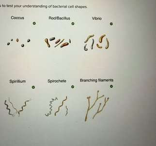
Coccus: Spherical bacteria
Bacillus: Rod-shaped bacteria
Vibrio: Comma-shaped bacteria
Spirillum: Spiral-shaped bacteria
Spirochete: Flexible, corkscrew-shaped bacteria
Branching Filaments: Bacteria with branching, filamentous structures
Step-by-Step Guidance
Review the main bacterial shapes: coccus, bacillus, vibrio, spirillum, spirochete, and branching filaments.
Understand how these shapes can be arranged (e.g., chains, clusters, pairs).
Be able to match images or diagrams to the correct shape and arrangement.
Practice labeling diagrams or concept maps with the correct terms.
Try solving on your own before revealing the answer!
Final Answer:
Bacterial shapes include coccus (spherical), bacillus (rod), vibrio (comma), spirillum (spiral), spirochete (corkscrew), and branching filaments. Arrangements can include chains, clusters, and pairs.
Q7. Simple Staining and Negative Staining
Background
Topic: Bacterial Staining Techniques
This question tests your understanding of simple staining, the difference between positive and negative stains, and the steps involved in preparing a bacterial smear.
Key Terms:
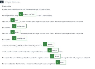
Simple Staining: Uses one dye to color cells, increasing contrast.
Positive Stain: Basic dye stains the cell, making it appear darker than the background.
Negative Stain: Acidic dye stains the background, leaving cells clear or lightly colored.
Bacterial Smear: A thin layer of bacteria spread on a slide, air-dried, and heat-fixed.
Step-by-Step Guidance
Understand the purpose of staining: to increase contrast and visualize cell shape, size, and arrangement.
Know the difference between basic (positive) and acidic (negative) stains.
Review the steps for preparing a bacterial smear: apply bacteria to slide, air dry, and heat fix.
Be able to identify images of positive and negative stains under the microscope.
Try solving on your own before revealing the answer!
Final Answer:
Simple staining uses one dye; positive stains color the cells, negative stains color the background. Bacterial smears are prepared by spreading bacteria, air drying, and heat fixing before staining.
Q8. Gram Staining: Steps and Interpretation
Background
Topic: Differential Staining - Gram Stain
This question tests your knowledge of the Gram staining procedure, the function of each reagent, and how to interpret results.
Key Terms and Steps:
Primary Stain (Crystal Violet): Colors all cells purple.
Mordant (Iodine): Fixes the dye inside the cell.
Decolorizer (Alcohol/Acetone): Removes dye from Gram-negative cells.
Counterstain (Safranin): Colors Gram-negative cells pink/red.
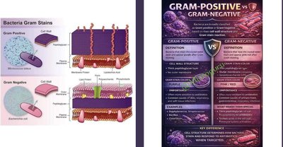
Gram-Positive: Retain purple color.
Gram-Negative: Appear pink/red after counterstaining.
Step-by-Step Guidance
List the steps of Gram staining in order: crystal violet, iodine, decolorizer, safranin.
Understand the function of each reagent in the process.
Know how to interpret the results: Gram-positive cells are purple, Gram-negative cells are pink/red.
Be able to match images of stained bacteria to their Gram reaction and cell wall structure.
Try solving on your own before revealing the answer!
Final Answer:
The Gram stain involves crystal violet (primary stain), iodine (mordant), alcohol/acetone (decolorizer), and safranin (counterstain). Gram-positive cells remain purple; Gram-negative cells turn pink/red.
Q9. Endospore Staining and Bacterial Genera
Background
Topic: Endospore Staining and Bacterial Pathogens
This question tests your understanding of endospore formation, the staining process, and diseases caused by endospore-forming bacteria.
Key Terms:
Endospore: A resistant, dormant structure formed by some bacteria under stress.
Bacillus and Clostridium: Genera of bacteria that form endospores.
Diseases: Anthrax, tetanus, botulism, gas gangrene.
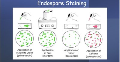
Staining Steps: Malachite green (primary), heat (mordant), water (decolorizer), safranin (counterstain).
Step-by-Step Guidance
Understand why bacteria form endospores (response to stress).
Know the genera that form endospores and the diseases they cause.
Review the steps of endospore staining and the function of each reagent.
Be able to identify endospores in stained preparations.
Try solving on your own before revealing the answer!
Final Answer:
Bacillus and Clostridium form endospores. Diseases include anthrax, tetanus, botulism, and gas gangrene. Endospore staining uses malachite green, heat, water, and safranin.
Q10. Selective and Differential Media: Mannitol Salt Agar and MacConkey Agar
Background
Topic: Microbial Growth Media
This question tests your understanding of how selective and differential media work, specifically Mannitol Salt Agar (MSA) and MacConkey Agar.
Key Terms:
Selective Media: Inhibits growth of some organisms while allowing others to grow.
Differential Media: Distinguishes between organisms based on biochemical reactions.
MSA: Selects for halophiles, differentiates mannitol fermenters (yellow color).
MacConkey: Selects for Gram-negative, differentiates lactose fermenters (pink color).

Step-by-Step Guidance
Understand what makes a medium selective or differential.
Know what MSA and MacConkey agar select for and how they differentiate bacteria.
Be able to interpret color changes on these media and what they indicate about the bacteria.
Practice explaining the purpose of each ingredient in the media.
Try solving on your own before revealing the answer!
Final Answer:
MSA selects for salt-tolerant bacteria and differentiates mannitol fermenters (yellow). MacConkey selects for Gram-negative bacteria and differentiates lactose fermenters (pink).
Q11. Viruses: Structure, Genome, and Classification
Background
Topic: Virology
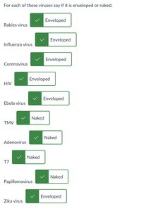
This question tests your knowledge of virus structure (enveloped vs. naked), genome types, and classification based on host and disease.
Key Terms:
Enveloped Virus: Has a lipid membrane surrounding the capsid.
Naked Virus: Lacks a lipid envelope.
Genome Types: ssRNA (single-stranded RNA), dsDNA (double-stranded DNA), segmented/non-segmented, linear/circular.
Host Range: The types of organisms a virus can infect.
Step-by-Step Guidance
Identify whether a virus is enveloped or naked based on its structure.
Classify viruses by their genome type (e.g., ssRNA, dsDNA, segmented, linear, circular).
Match viruses to their host range and associated diseases.
Understand the significance of these features for viral replication and pathogenesis.
Try solving on your own before revealing the answer!
Final Answer:
Viruses can be enveloped or naked, with genomes of ssRNA, dsDNA, etc. Classification depends on structure, genome, and host range. Examples: Influenza (enveloped, ssRNA, segmented), Adenovirus (naked, dsDNA).
