 Back
BackMicroscopy in Microbiology: Principles, Techniques, and Staining Methods
Study Guide - Smart Notes
 Tailored notes based on your materials, expanded with key definitions, examples, and context.
Microscopes and Measurement in Microbiology
Units of Measurement
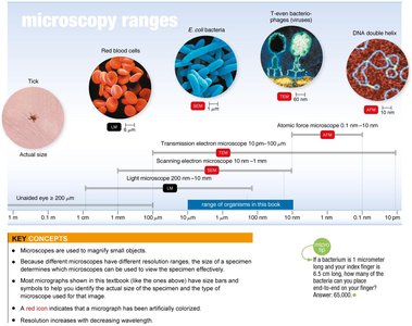
Microorganisms are extremely small and require specialized units for measurement. The most common units are micrometers (μm) and nanometers (nm).
Micrometer (μm): 1 μm = 10-6 meters
Nanometer (nm): 1 nm = 10-9 meters
1000 nm = 1 μm
1 μm = 1000 nm
Example: The average size of a bacterium is about 1 μm.
Principles of Microscopy
Types of Microscopes
Microscopes are essential tools in microbiology, allowing visualization of organisms too small to be seen with the naked eye. There are several types of microscopes, each with unique features and applications.
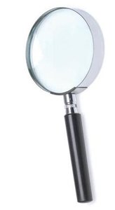
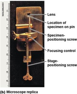
Simple Microscope: Contains a single lens, similar to a magnifying glass but with higher magnification.
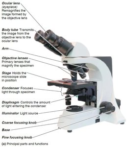
Compound Light Microscope: Uses multiple lenses and visible light to magnify specimens.
Magnification and Resolution
Magnification is the process of enlarging the appearance of an object. In a compound microscope, total magnification is calculated as:
Ocular lens is typically 10X.
Objective lenses commonly include 4X, 10X, 40X, and 100X.
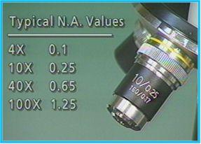
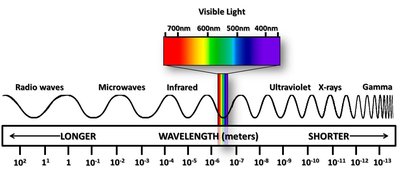
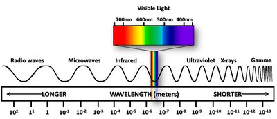
Resolution is the ability of a lens to distinguish two points as separate entities. Higher resolution allows for clearer, more detailed images. Resolution increases as the wavelength of light used decreases.
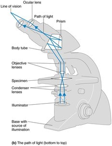
Path of Light in a Compound Microscope
Light passes through several components in a compound microscope:
Illuminator (light source)
Condenser lens
Specimen
Objective lens
Body tube (with prism)
Ocular lens (eyepiece)
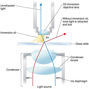
Oil Immersion Technique
Oil immersion is used with the 100X objective lens to increase resolution. Immersion oil has a refractive index similar to glass, reducing light refraction and allowing more light to enter the objective lens.
Types of Light Microscopy
Brightfield Microscopy
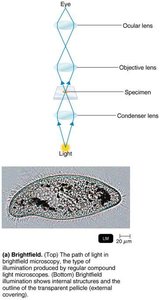
Brightfield microscopy is the most common form, where dark objects are visible against a bright background. It is useful for stained specimens and reveals internal structures.
Darkfield Microscopy
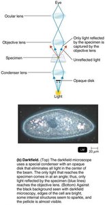
Darkfield microscopy uses a special condenser with an opaque disk, making light objects visible against a dark background. It is ideal for observing live, unstained specimens, such as spirochetes.
Phase-Contrast Microscopy
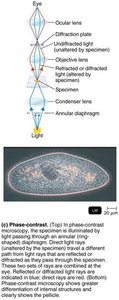
Phase-contrast microscopy enhances contrast in transparent specimens without staining. It brings together direct and diffracted light rays to visualize internal cell structures in living cells.
Differential Interference Contrast (DIC) Microscopy
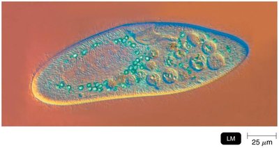
DIC microscopy is similar to phase-contrast but uses two light beams and prisms to produce high-contrast, colorful images with a three-dimensional appearance.
Fluorescence Microscopy
Fluorescence microscopy uses ultraviolet (UV) light to excite fluorescent dyes (fluorochromes) in specimens, causing them to emit visible light. It is widely used for detecting specific microbes using fluorescent antibodies.
Confocal Microscopy
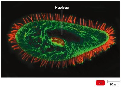
Confocal microscopy uses fluorochrome dyes and a laser to scan specimens in thin planes, creating detailed three-dimensional images. It is useful for examining thick specimens and biofilms.
Two-Photon Microscopy
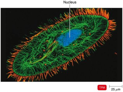
Two-photon microscopy uses long-wavelength (red) light to excite fluorochromes, allowing imaging of living cells up to 1 mm deep. It is valuable for studying cell interactions in tissues.
Super-Resolution Light Microscopy
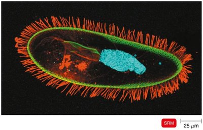
Super-resolution microscopy uses two laser beams to achieve resolution beyond the diffraction limit of light, allowing visualization of structures at the nanometer scale.
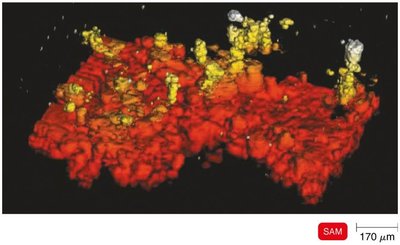
Scanning Acoustic Microscopy (SAM)
SAM uses sound waves to study cells attached to surfaces, with a resolution of about 1 μm. It is useful for examining biofilms and surface structures.
Electron and Probe Microscopy
Electron Microscopy
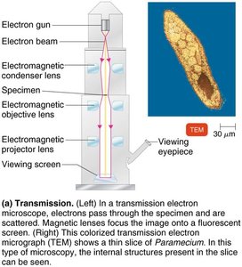
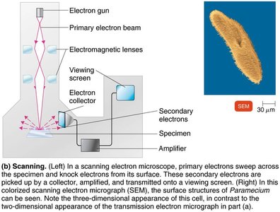
Electron microscopes use electron beams instead of light, providing much higher resolution. They require a vacuum and cannot be used for live specimens.
Transmission Electron Microscopy (TEM): Electrons pass through ultrathin sections of specimens, revealing internal structures. Magnification up to 10,000,000x; resolution of 10 pm.
Scanning Electron Microscopy (SEM): Electrons scan the surface, producing detailed three-dimensional images of specimen surfaces. Magnification up to 500,000x; resolution of 10 nm.
Scanning Probe Microscopy
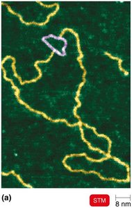
Scanning Tunneling Microscopy (STM): Uses a tungsten probe to scan surfaces at atomic resolution (1/100 of an atom).
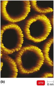
Atomic Force Microscopy (AFM): Uses a metal-and-diamond probe to produce three-dimensional images at near-atomic detail.
Staining Techniques in Microbiology
Preparation of Specimens
Staining enhances contrast in microscopic images. A smear is a thin film of microorganisms spread on a slide and fixed by heat to kill and attach the cells.
Basic dyes: Chromophore is a cation (+), stains the cell.
Acidic dyes: Chromophore is an anion (−), stains the background (negative staining).
Simple Staining
Simple stains use a single basic dye to highlight the entire microorganism, making cell shapes and structures visible. A mordant may be used to intensify the stain.
Differential Staining
Differential stains distinguish between different groups of bacteria. The two main types are the Gram stain and the acid-fast stain.
Gram Stain
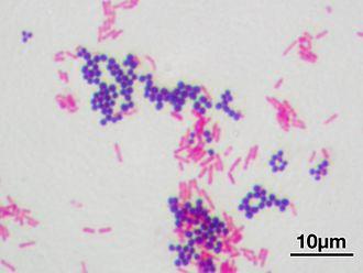
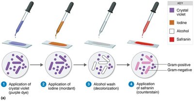
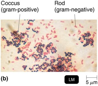
The Gram stain classifies bacteria as gram-positive or gram-negative based on cell wall structure.
Gram-positive: Thick peptidoglycan cell wall (stains purple/blue).
Gram-negative: Thin peptidoglycan and outer membrane (stains pink/red).
Step | Gram-Positive | Gram-Negative |
|---|---|---|
Crystal violet (primary stain) | Purple | Purple |
Iodine (mordant) | Purple | Purple |
Alcohol (decolorizer) | Purple | Colorless |
Safranin (counterstain) | Purple | Pink |
Acid-Fast Stain
The acid-fast stain identifies bacteria with waxy cell walls (mycolic acid), such as Mycobacterium and Nocardia. Acid-fast bacteria retain the primary stain (carbolfuchsin) after acid-alcohol decolorization and appear red; non–acid-fast bacteria appear blue after counterstaining with methylene blue.
Step | Acid-Fast Bacteria | Non–Acid-Fast Bacteria |
|---|---|---|
Carbolfuchsin (primary stain) | Red | Red |
Acid-alcohol (decolorizer) | Red | Colorless |
Methylene blue (counterstain) | Red | Blue |
Special Stains
Special stains are used to highlight specific structures:
Capsule stain: Negative staining to visualize gelatinous capsules as halos around cells.
Endospore stain: Endospores are stained with malachite green (with heat), while the rest of the cell is counterstained with safranin. Endospores appear green within red or pink cells.
Flagella stain: Uses a mordant and carbolfuchsin to thicken flagella, making them visible under the microscope.
Summary Table: Types of Microscopy and Their Applications
Microscopy Type | Principle | Application |
|---|---|---|
Brightfield | Light passes through specimen | Stained cells, general morphology |
Darkfield | Light reflected by specimen | Live, unstained cells (e.g., spirochetes) |
Phase-Contrast | Combines direct and diffracted light | Internal structures of living cells |
DIC | Two beams, prisms for color/contrast | 3D, colorful images of cells |
Fluorescence | UV light excites fluorochromes | Detecting specific microbes |
Confocal | Laser scans stained specimen | 3D images, biofilms |
Electron (TEM/SEM) | Electron beams | Ultrastructure, viruses, surfaces |
Scanning Probe (STM/AFM) | Physical probe scans surface | Atomic/molecular detail |
Additional info: The choice of microscopy and staining technique depends on the research question, the type of microorganism, and the structures of interest. Proper specimen preparation is crucial for accurate observation and identification in microbiology.
