 Back
BackMicroscopy in Microbiology: Principles, Types, and Applications
Study Guide - Smart Notes
 Tailored notes based on your materials, expanded with key definitions, examples, and context.
Microscopy: Observing Microorganisms
Units of Measurement
Microorganisms are measured using the metric system, primarily in micrometers (µm) and nanometers (nm). Understanding these units is essential for interpreting microscopic observations.
Micrometer (µm): 1 µm = 10-6 meters
Nanometer (nm): 1 nm = 10-9 meters
1 µm = 1000 nm
Example: Most bacteria are 1–10 µm in length, while viruses range from 20–300 nm.
Types of Microscopes
Microscopes are essential tools in microbiology, allowing visualization of structures too small for the naked eye. The two main categories are light microscopes and electron microscopes.
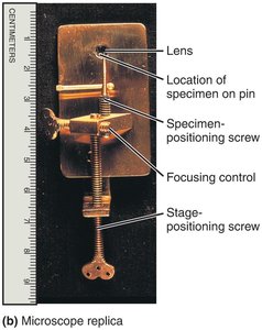
Simple Microscope
A simple microscope uses a single lens for magnification, similar to a magnifying glass but with higher quality optics. Anton van Leeuwenhoek's early observations of microorganisms were made with such an instrument.
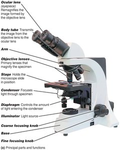
Compound Light Microscope
The compound light microscope uses multiple lenses to achieve higher magnification and resolution. It is the standard instrument in microbiology labs.
Ocular lens (eyepiece): Remagnifies the image formed by the objective lens
Objective lenses: Primary lenses that magnify the specimen (commonly 4x, 10x, 40x, 100x)
Stage: Holds the microscope slide
Condenser: Focuses light through the specimen
Diaphragm: Controls the amount of light entering the condenser
Coarse and fine focusing knobs: Adjust the focus
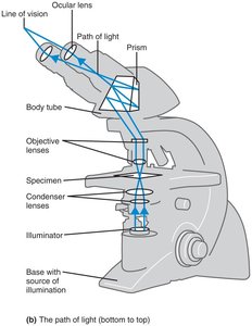
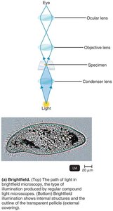
Path of Light in a Compound Microscope
Light from the illuminator passes through the condenser, specimen, objective lens, and ocular lens, forming a magnified image for the observer.
Total Magnification and Resolution
Total Magnification: Calculated by multiplying the magnification of the objective lens by that of the ocular lens.
Resolution (Resolving Power): The ability to distinguish two points as separate entities. Higher resolution allows for clearer, more detailed images. The limit of resolution for a compound light microscope is about 0.2 µm.
Wavelength: Shorter wavelengths of light provide greater resolution.
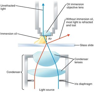
Refractive Index and Immersion Oil
The refractive index is a measure of how much a medium bends light. Immersion oil is used with high-power objectives to reduce light refraction and increase resolution.
Types of Light Microscopy
Brightfield Microscopy
Brightfield microscopy is the most common form, where dark objects are visible against a bright background. It is suitable for stained specimens but may lack contrast for live, unstained cells.
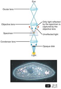
Darkfield Microscopy
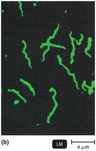
Darkfield microscopy enhances the contrast of unstained, live specimens. Only light reflected by the specimen enters the objective lens, making the specimen appear bright against a dark background. Useful for observing thin organisms like Treponema pallidum.
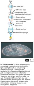
Phase-Contrast Microscopy
Phase-contrast microscopy allows detailed examination of living cells and internal structures without staining. It uses differences in refractive index to produce high-contrast images of transparent specimens.
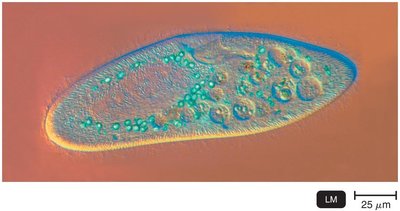
Differential Interference Contrast (DIC) Microscopy
DIC microscopy uses two beams of light and prisms to produce high-contrast, brightly colored, three-dimensional images of live specimens.
Fluorescence Microscopy
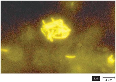
Fluorescence microscopy uses ultraviolet (UV) light to excite fluorescent dyes (fluorochromes) that emit visible light. It is widely used for detecting specific microbes using fluorescent-antibody techniques (immunofluorescence).
Auramine O: Stains Mycobacterium tuberculosis bright yellow
Immunofluorescence: Uses antibodies tagged with fluorochromes for rapid pathogen detection
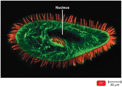
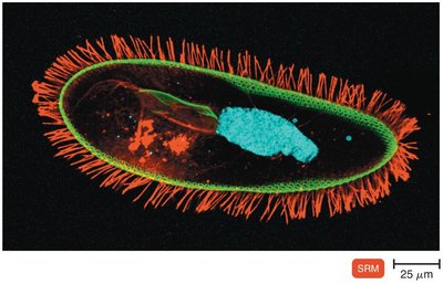
Confocal Microscopy
Confocal microscopy uses lasers and fluorochromes to obtain sharp, two-dimensional images at various depths, which can be reconstructed into three-dimensional images. It is valuable for studying complex structures in cells and biofilms.
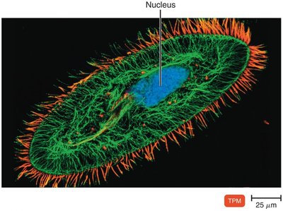
Two-Photon Microscopy
Two-photon microscopy uses long-wavelength (red) light to excite fluorochromes, allowing imaging of living cells up to 1 mm deep and tracking cellular activity in real time.
Super-Resolution Light Microscopy
Super-resolution microscopy surpasses the diffraction limit of light, enabling visualization of structures at the nanometer scale. It uses advanced laser techniques and computational reconstruction for single-molecule tracking and high-resolution imaging.
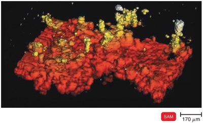
Scanning Acoustic Microscopy (SAM)
SAM uses sound waves reflected from a specimen to generate images. It is useful for studying cells attached to surfaces, such as biofilms, with a resolution of about 1 µm.
Electron Microscopy
Principles of Electron Microscopy
Electron microscopes use electron beams instead of light, providing much higher resolution and magnification. They are essential for visualizing viruses and internal cellular structures.
Transmission Electron Microscope (TEM): Electrons pass through ultrathin sections of specimens, revealing internal structures. Magnification: 10,000–10,000,000x; resolution: 0.2 nm.
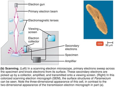
Scanning Electron Microscope (SEM): Electrons scan the surface of specimens, producing detailed three-dimensional images. Magnification: 1,000–500,000x; resolution: 0.5 nm.

Scanned-Probe Microscopy
Scanned-probe microscopes use physical probes to scan specimen surfaces, providing atomic or near-atomic resolution without specimen modification.
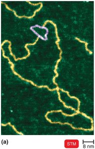
Scanning Tunneling Microscopy (STM): Uses a tungsten probe to scan surfaces, resolving features as small as atoms. No special preparation is needed.
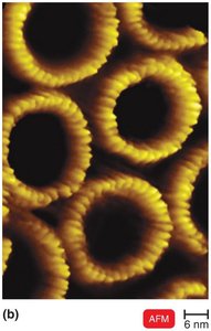
Atomic Force Microscopy (AFM): Uses a metal-and-diamond probe to produce three-dimensional images at near-atomic detail.
Staining and Preparing Microbial Specimens
Staining Techniques
Staining enhances contrast in microscopic images by coloring microorganisms or their background. Fixation (by heat or chemicals) attaches and preserves cells on slides.
Basic dyes: Chromophore is a cation (e.g., crystal violet, methylene blue, safranin); stains bacterial cells directly.
Acidic dyes: Chromophore is an anion (e.g., eosin, acid fuchsin, nigrosin); stains the background (negative staining).
Simple Staining
Simple stains use a single basic dye to highlight the entire microorganism, making cell shapes and structures visible. A mordant may be used to intensify the stain.

Differential Staining
Differential stains distinguish between different types of bacteria or structures. The most important are the Gram stain and the acid-fast stain.
Gram Stain
The Gram stain classifies bacteria as gram-positive (thick peptidoglycan, purple) or gram-negative (thin peptidoglycan, outer membrane, pink/red). It is crucial for bacterial identification and treatment decisions.
Acid-Fast Stain
The acid-fast stain identifies bacteria with waxy cell walls (e.g., Mycobacterium, Nocardia). Acid-fast cells retain the primary stain (red) after acid-alcohol decolorization; non–acid-fast cells take up the counterstain (blue).
Special Stains
Special stains highlight specific microbial structures:
Capsule stain: Visualizes the gelatinous capsule surrounding some bacteria.
Endospore stain: Detects resistant spores within bacteria.
Flagella stain: Reveals bacterial flagella for motility studies.
Summary Table: Types of Microscopy
Microscopy Type | Principle | Best Use | Resolution |
|---|---|---|---|
Brightfield | Light passes through specimen | Stained cells | 0.2 µm |
Darkfield | Only reflected light enters lens | Live, unstained cells | 0.2 µm |
Phase-Contrast | Exploits differences in refractive index | Internal structures of live cells | 0.2 µm |
DIC | Two beams, prisms for 3D effect | Live, unstained cells | 0.2 µm |
Fluorescence | UV light excites fluorochromes | Specific detection of microbes | 0.2 µm |
Confocal | Laser scans planes, 3D images | Thick specimens, biofilms | 0.2 µm |
Electron (TEM) | Electrons pass through specimen | Internal structures, viruses | 0.2 nm |
Electron (SEM) | Electrons scan surface | Surface details, 3D images | 0.5 nm |
STM/AFM | Physical probe scans surface | Atomic/molecular detail | ~0.1 nm |
Additional info: This guide covers the foundational concepts and practical applications of microscopy in microbiology, including measurement units, microscope types, principles of magnification and resolution, and essential staining techniques for observing microorganisms.
