 Back
BackMicroscopy in Microbiology: Tools, Types, and Applications
Study Guide - Smart Notes
 Tailored notes based on your materials, expanded with key definitions, examples, and context.
Microscopy in Microbiology
Introduction to Microscopy
Microscopy is fundamental to microbiology, enabling the visualization and study of microorganisms that are otherwise invisible to the naked eye. The invention and continual improvement of microscopes have driven major advances in the field, allowing scientists to observe, describe, and understand microbial life.
Microscope: An instrument that magnifies small objects, making them visible and analyzable.
Microbiology: The study of microscopic organisms, including bacteria, viruses, fungi, and protozoa.
Historical Impact: Microbiology as a science began with the invention of the microscope.
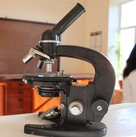
Discovery of Microorganisms
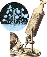
The existence of microorganisms was suspected for centuries, but direct observation was only possible after the development of microscopes. Early pioneers made significant contributions to the discovery and description of microbes.
Robert Hooke (1635–1703): Published Micrographia (1665), the first book of microscope observations, including drawings of mold structures.
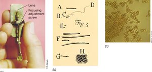
Antoni van Leeuwenhoek (1632–1723): First to observe bacteria using single-lens light microscopes, reporting his findings in 1676.
Magnification: Leeuwenhoek's microscopes could magnify up to ~266×.
Types of Microscopes
Light Microscopy
Light microscopy (optical microscopy) uses visible light and optical lenses to magnify specimens. It is widely used in biology for observing living cells and tissues, offering real-time visualization and a range of contrast techniques.
Principle: Light is shone on or through a specimen, and lenses focus and enlarge the image.
Observation: The image is viewed through an eyepiece or recorded with a camera.
Common Types: Brightfield, fluorescence, phase contrast, and stereo (dissection) microscopes.
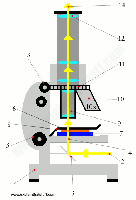
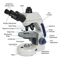
Brightfield (Compound) Microscopy
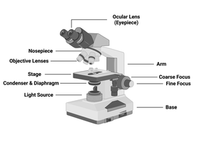
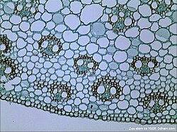
Brightfield microscopy is the most common form of light microscopy. It uses visible light to illuminate a sample, which appears darker against a bright background. This technique is best for stained samples but offers low contrast for transparent specimens.
Main Parts: Light source, objective lenses, eyepiece/camera, stage, diaphragm, focusing knobs.
Sample Preparation: Staining is often required to enhance contrast.
Drawback: Transparent or unstained samples are difficult to see.
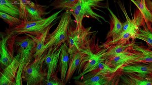
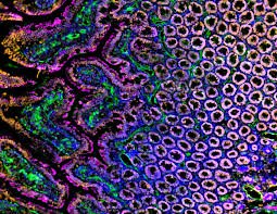
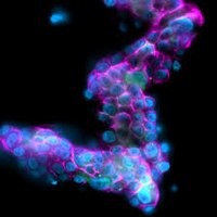
Fluorescence Microscopy
Fluorescence microscopy uses high-energy light and fluorescent dyes (fluorophores) to highlight specific cellular components. It provides high-contrast images and reveals specific structures and processes that are invisible in regular light microscopy.
Principle: Fluorophores absorb excitation light and emit lower-energy light (glow), which is filtered and visualized.
Light Sources: Lamps, LEDs, or lasers.
Applications: Medical diagnostics, cell biology, neuroscience, drug discovery.
Phase Contrast Microscopy
Phase contrast microscopy enhances the visibility of unstained, living cells by converting phase shifts in light passing through the specimen into differences in brightness. This technique allows for detailed observation of internal structures without the need for staining.
Inventor: Frits Zernike (1930s), Nobel Prize in Physics (1953).
Advantage: Non-toxic, real-time observation of living cells.
Stereomicroscope (Dissection Microscope)
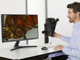
Stereomicroscopes provide a three-dimensional view of larger specimens at low magnification. They are ideal for examining insects, plants, fossils, or circuit boards, offering a large working space and upright images.
Magnification: Typically 5×–50×.
3D View: Two light paths for depth perception.
Applications: Dissection, manipulation, and inspection of macroscopic samples.
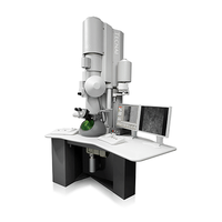
Electron Microscopy
Principles and Applications
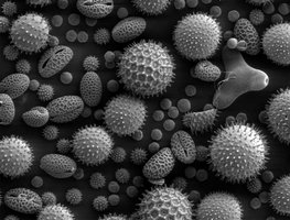
Electron microscopy (EM) uses a beam of electrons instead of light to achieve extremely high magnification and resolution. Electrons have much shorter wavelengths than visible light, allowing for detailed imaging of cellular and molecular structures.
Principle: Electron gun generates electrons, electromagnetic lenses focus the beam, and signals are detected to form an image.
Environment: EM operates in a vacuum to prevent electron scattering.
Applications: Study of cell ultrastructure, viruses, and nanomaterials.
Summary Table: Types of Microscopes
Type | Principle | Magnification | Best For |
|---|---|---|---|
Brightfield | Visible light, stained samples | Up to 1000× | Bacteria, cells, tissues |
Fluorescence | Fluorophores, high-energy light | Up to 1000× | Cell components, diagnostics |
Phase Contrast | Phase shifts, contrast enhancement | Up to 1000× | Living, unstained cells |
Stereomicroscope | Two light paths, 3D view | 5×–50× | Large specimens, dissection |
Electron Microscope | Electron beam, electromagnetic lenses | Up to 1,000,000× | Cell ultrastructure, viruses |
Key Equations
Magnification:
Resolution: where is the wavelength of light/electrons, and NA is the numerical aperture of the lens.
Conclusion
Microscopy is indispensable in microbiology, providing the means to observe, identify, and analyze microorganisms. Understanding the principles, types, and applications of microscopes is essential for any microbiology student.
